Chest Imaging in COVID-19 #Thoracic Radiology #Covid-19 #Lungs

Introduction:
A pandemic flu is different from the seasonal flu in many ways. Seasonal flu strikes annually and usually people have immunity to a variable extent due to past exposures. Only people with risk factors are more susceptible. Pandemic flu, on the other hand, is rare (H1N1 in 1918) and people do not have any immunity to the virus. As a result, even healthy young adults can get infected. The common denominator between the two is rapid spread through human to human transmission, thereby having pandemic potential.
Epidemiology and Risk factors:
The current Covid-19 pandemic has spanned across 215 countries and territories and affected more than 4.4 million people with close to 300 thousand (3 Lakhs) deaths, at the time of writing this article.
The spread of infection is through droplets while coughing/ sneezing/ talking and aerosol generating procedures such as intubation. The virus is also known to stay on surfaces which have droplets and is transmitted to others when touched by hands and, the hands, in turn, touch the nose, mouth or eyes.
In a nationwide study in China of over 1590 hospitalized patients, risk assessment was done to determine their prognosis. They concluded that targeting the at-risk patients earlier, intensive surveillance and early treatment measures, are the best way forward. The most important independent risk factor is advanced age, with the risk being almost 7 times more for those aged above 75 years. Associated chronic illnesses such as coronary heart disease and cerebrovascular disease are strong independent risk factors as well. Dyspnea at presentation carries high risk due to patients progressing to complication such as ARDS. Laboratory abnormalities such as raised PCT (> 0.5 ng/ml) (produced from C cells of thyroid gland) indicate bacterial infection or sepsis and carry poor prognosis. Elevated serum creatinine (133 µmol/L), AST (> 40 U/L) and total bilirubin (> 17.1 µmol/L) are associated with fatal outcome as well. Other risk factors include malignancy, chronic renal disease, diabetes, hypertension, obesity (BMI > 40) and hepatitis B infection (1).
Chest Imaging Protocols:
There are two modalities, namely, Chest X Ray and CT scan, that have been recommended for imaging. Ultrasound use is not recommended due to its lack of sensitivity and specificity, operator dependence and risk of exposure to healthcare workers. The recommendations from Fleischner Society in a Multinational Consensus Statement are summarized as 3 clinical scenarios (2).
- Imaging is not a screening test. Begin testing for suspected Covid-19 with RT-PCR. Imaging is to be used for patients with mild symptoms ONLY if they have associated risk factors for disease progression. This includes chronic illnesses, obesity etc. listed above. The pre-test probability of disease (disease prevalence) is not of concern.
- Imaging is to be used for all suspected Covid-19 patients with moderate to severe symptoms, irrespective of Covid-19 status by RT-PCR or pre-test probability of disease.
- Imaging is used in Triage of patients with moderate to severe respiratory symptoms of Covid-19, once the resources are overwhelmed in a region with high pre-test probability (and disease prevalence). This is to be adopted when the number of ICU beds, ventilators and other resources are in short supply and critical decisions for admissions need to be carried out.
Additional Guidelines:
- In stable intubated patients with Covid-19, daily imaging is not indicated.
- In patients recovered from Covid-19, a functional impairments or hypoxemia warrants CT imaging. This helps in prognosis and to look for other etiologies such as secondary infections or thromboembolism.
- Covid-19 testing with RT-PCR is indicated in patients with incidental lung findings of Covid-19 on CT scans performed for other indications (such as on abdomen CT).
Although, a wide permutation of scenarios are clinically possible in real life depending on resources and situations, in which case, the Radiologist takes a call on imaging based on proactive multidisciplinary discussions.

Chest Imaging Findings:
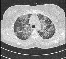
Classical Covid-19 chest imaging findings, as observed consistently across the affected countries are, bilateral, peripheral and lower lobe distribution, rounded ground-glass opacities. Crazy paving appearance and a reverse atoll (reverse halo) sign are well documented as well. Air bronchogram, air trapping, vascular enlargement and interlobular septal thickening are seen at initial CT related to lung injury and edema (3).
However, there are certain features which should make one think of alternate diagnosis: 1) Lymphadenopathy, 2) Cavitation, 3) Pleural effusion, 4) Nodules (especially tree-in-bud or centrilobular) and 5) Isolated lobar or segmental consolidation.

Figure 1: Bilateral lower lobe predominant rounded ground-glass opacities, in a Covid-19 patient. [Radiology. 2020 Feb 20:200463]

Figure 2: Bilateral lower lobe predominant peripheral ground-glass opacities and subsegmental consolidation, in a Covid-19 patient. [Radiology. 2020 Feb 20:200463]
On Follow up CT scans, performed between 2-15 days after the initial CT scan, disease worsening is indicated by increase in number and size of ground-glass opacities or consolidation, involvement of greater number of lobes or ‘white out lung’ suggesting acute respiratory distress syndrome (ARDS) and carry poor prognosis. Disease regression is indicated by appearance of reticular opacities and fibrosis or resolution of ground-glass opacities or consolidation and carry better prognosis. Although, the fibrosis and distortion of bronchi may cause some permanent impairment in lung function (3).
Chest Imaging Covid-19 Reporting Templates:
The Covid-19 Chest Imaging reporting templates have been standardized by the Radiological Society of North America Expert Consensus Statement and is endorsed by the Society of Thoracic Radiology (STR), the American College of Radiology (ACR), and Radiological Society of North America (RSNA). After reporting the Chest Imaging findings in observations, with emphasis on the Covid-19 pertaining features, the Impression of the Report is to include one of the following categories. The templates are divided into four categories on CT Chest Without Contrast (4):
- Typical for Covid-19: Bilateral peripheral, multifocal, rounded ground glass opacities or crazy-paving in lower lobe predominant distribution on imaging.
- Indeterminate for Covid-19: Bilateral multifocal non-rounded ground glass opacities in central distribution with no lower lobe predominance. In these cases, features could represent infection (such as viral pneumonias) or inflammatory etiologies, drug toxicity etc.
- Atypical for Covid-19: Bilateral multiple tree-in-bud or centrilobular nodules or unilateral single lobar or segmental consolidation or cavitation or lymphadenopathy or pleural effusion or isolated smooth interlobular septal thickening. In these cases, alternative diagnosis should be considered.
- Negative for Covid-19: No abnormal features on CT Chest.
In our hospital, each Typical and Indeterminate report is documented as ‘critical’ and reported as “Critical findings communicated to Dr. xyz on [date] at [time] over mail/phone”. The referring doctor also receives a mandatory call from the Radiologist to apprise them of the relevant findings, in these cases.
In areas where disease prevalance is low, the above reporting template works well. This is especially true when RT-PCR is unavailable and Covid status is unknown.
In areas where disease prevalance is high, the reporting templates can be more standardized to help in decision making based on cut off points. This is true for RT-PCR positive patients or known Covid patients. The table below gives the reporting template categories in this scenario, for the level of suspicion of pulmonary Covid involvement.
CO-RADS Category | Level of suspicion for pulmonary involvement of Covid-19 | Summary |
CO-RADS 0 | Not interpretable | scan technically insufficient for assigning a score |
CO-RADS 1 | Very low | normal or non-infectious |
CO-RADS 2 | Low | typical for other infection but not COVID-19 |
CO-RADS 3 | Equivocal/ unsure | features compatible with COVID-19, but also other diseases |
CO-RADS 4 | High | suspicious for COVID-19 |
CO-RADS 5 | Very high | typical for COVID-19 |
CO-RADS 6 | Proven | RT-PCR positive SARS-CoV-2 |
Table 1. Overview of CO-RADS categories and the level of suspicion of lung involvement in Covid-19 (5).

Radiology Department Protocols in Covid times:
The Radiology Society of North America (RSNA) has provided clearly laid out guidelines to help countries and communities facing or that are yet to face the full wrath of the pandemic. The various questions on how to prevent infection amongst other patients undergoing imaging, how to clean the room have been answered (6).
In general, a disinfectant based wipe down of the CT room and gantry is performed after scanning any suspected or positive Covid-19 patient. In situations when possible, a designated X-ray machine is allocated for imaging these patients with contract and droplet precautions. In addition, at Manipal Hospitals, a dedicated bedside ultrasound equipment is reserved for these patients.
The hospital staff including doctors, technologists, nurses and housekeeping are donning appropriate PPE depending on work requirement and proximity to suspected and positive Covid-19 patients. Social distancing and hand sanitization are followed at every level.
Pediatric Chest Imaging Findings:
The Chest Imaging findings in pediatrics follow the same pattern and reporting templates.
First Response Center:
In Bangalore, India, Manipal Hospitals at Yeswanthpur has been designated as the first responder in that region by the State Government. The entire group of Manipal Hospitals is prepared as per above guidelines, protocols and reporting templates with minor individual hospital specific changes.
Teleradiology:
Teleradiology section ofManipal Hospitals Radiology Group is prepared with above protocols and reporting templates and ready to serve its over 70 teleradiology sites, that might soon experience a surge in cases. At an individual level, every Radiologist and every doctor is committed to his/ her role and is updating themselves periodically as this is an evolving unusual situation.
Conclusion:
As we face the 2019-2020 Covid-19 pandemic, every hospital and all individuals associated, prepare in an energetic and organized way. The guidelines are followed top to bottom, so we direct our efforts and care in full force in a unified direction with appropriate Radiology department protocols, knowledge of chest imaging findings and reporting templates while following the dos and don’ts in a pandemic. This is a rapidly changing situation, where we should all be ready to adapt and implement at short notice.
References:
- Chen R, Liang W, Jiang M, Guan W, Zhan C, Wang T, Tang C, Sang L, Liu J, Ni Z, Hu Y. Risk factors of fatal outcome in hospitalized subjects with coronavirus disease 2019 from a nationwide analysis in China. Chest. 2020 Apr 15.
- Rubin GD, Ryerson CJ, Haramati LB, Sverzellati N, Kanne JP, Raoof S, Schluger NW, Volpi A, Yim JJ, Martin IB, Anderson DJ. The role of chest imaging in patient management during the COVID-19 pandemic: a multinational consensus statement from the Fleischner Society. Chest. 2020 Apr 7.
- Li Y, Xia L. Coronavirus disease 2019 (COVID-19): role of chest CT in diagnosis and management. American Journal of Roentgenology. 2020 Feb 21:1-7.
- Simpson S, Kay FU, Abbara S, Bhalla S, Chung JH, Chung M, Henry TS, Kanne JP, Kligerman S, Ko JP, Litt H. Radiological Society of North America Expert Consensus Statement on Reporting Chest CT Findings Related to COVID-19. Endorsed by the Society of Thoracic Radiology, the American College of Radiology, and RSNA. Radiology: Cardiothoracic Imaging. 2020 Mar 25;2(2):e200152.
- Prokop M, van Everdingen W, van Rees Vellinga T, Quarles van Ufford J, Stöger L, Beenen L, Geurts B, Gietema H, Krdzalic J, Schaefer-Prokop C, van Ginneken B. CO-RADS–A categorical CT assessment scheme for patients with suspected COVID-19: definition and evaluation. Radiology. 2020 Apr 27:201473.
- Mossa-Basha M, Meltzer CC, Kim DC, Tuite MJ, Kolli KP, Tan BS. Radiology department preparedness for COVID-19: radiology scientific expert panel. Radiology. 2020 Mar 16:200988.
Author
Dr Deepali Saxena MBBS, DNB, Cardiothoracic Imaging Fellowship (USA)
Senior Consultant Radiologist,
Lead Cardiac Imaging & Specialist Chest Imaging,
Manipal Hospitals Radiology Group