G2E1 with 37+5 weeks of gestation with PROM delivered male baby with antenatal scan showing
G2E1 with 37+5 weeks of gestation with PROM delivered male baby with antenatal scan showing
- Prominent small Bowel Loops
- ?Small Bowel Atresia
FINDINGS:
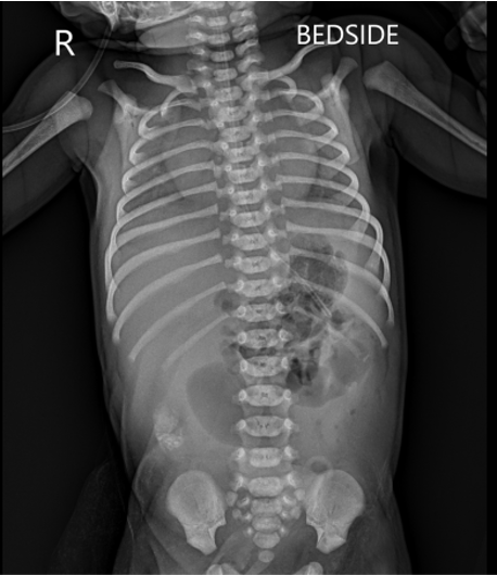
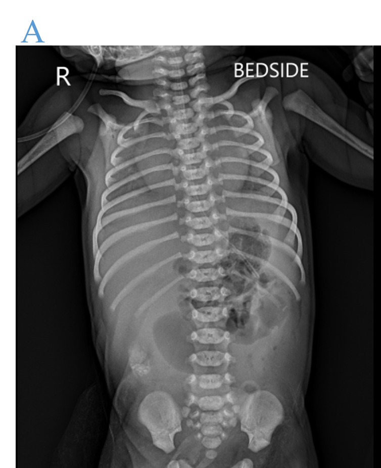
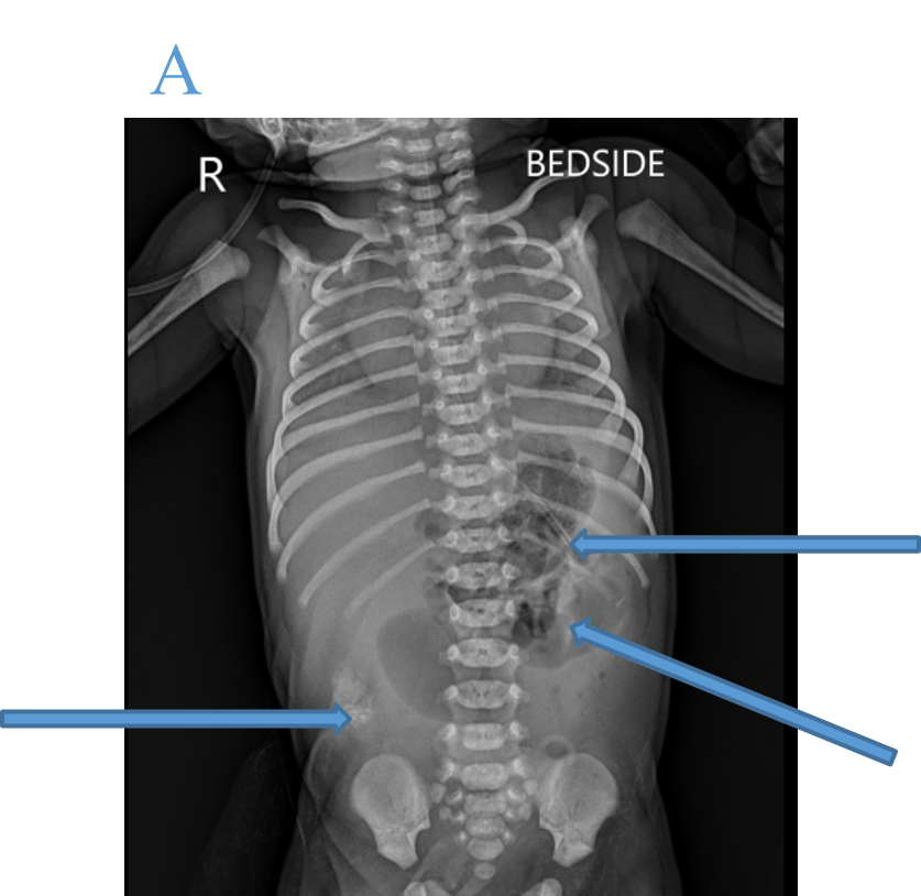
- X RAY ABDOMEN AP
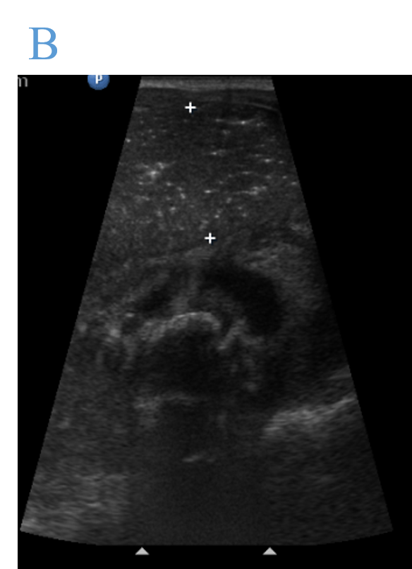
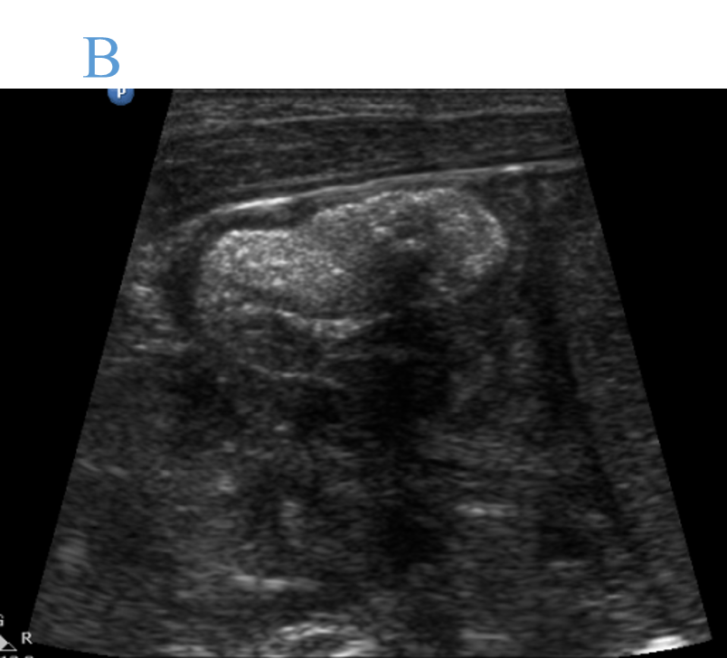
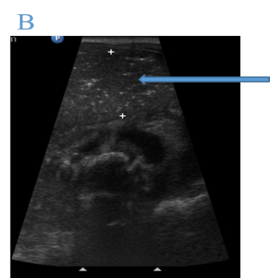
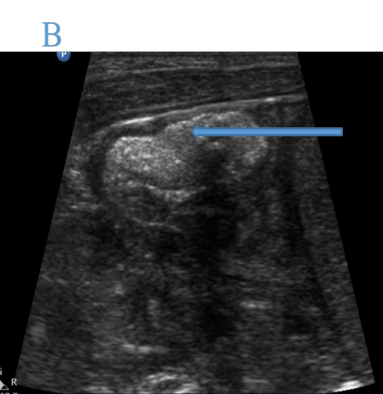
- USG ABDOMEN AND PELVIS
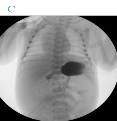
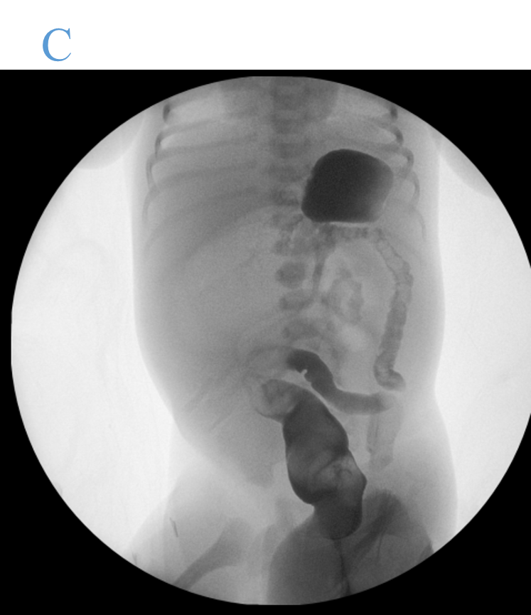
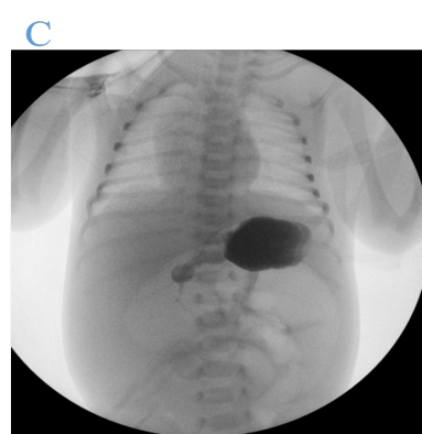
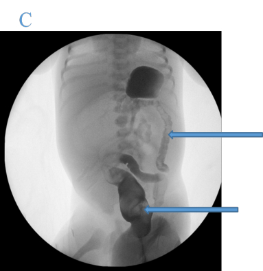
- UPPER AND LOWER GI CONTRAST STUDY
- Dilated air filled proximal small bowel loops with no evidence of air in distal loops
- High density meconium in right iliac fossa.
- Nasogastric tube tip in stomach.
- Fluid filled distension of stomach & dilated proximal small bowel loops.
- Intraluminal meconium in distal small bowel loops.
- Upper GI study –No evidence of malrotation.
- Lower GI study – Rectum filled with meconium, small caliber colon measuring 6 mm suggestive of microcolon
DIAGNOSIS:
- Mid-Distal Small Bowel Atresia and obstructi
TREATMENT:
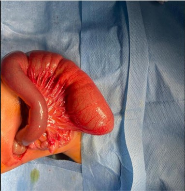
Right upper quadrant laparotomy
- Intestinal resection of jejunoileal atresia, end-back intestinal Anastomosis and intra-peritoneal drain insertion
POST OPERATIVE X RAY CHEST AP
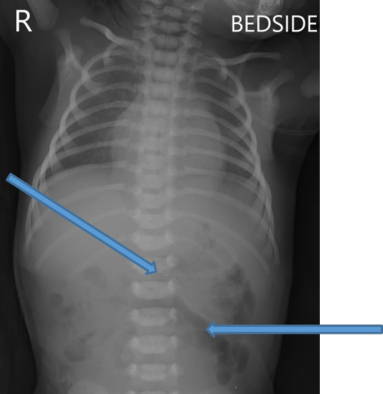
- Air noted in distal bowel loops.
- Previously noted high density meconium seen in right iliac fossa is noted seen – passed out meconium.
- Abdominal drain noted in central abdomen region.
HPE REPORT
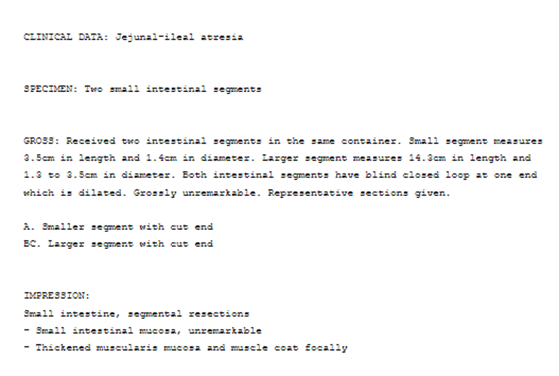
DISCUSSION:
Jejunal Atresia
- Jejunal atresia is due to an ischemic insult in utero.
- Neonates present early with vomiting, abdominal distention, and failure to pass meconium.
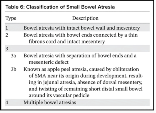
- Small bowel atresia is classified into four categories:
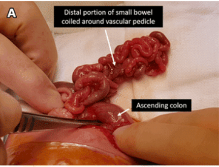
Apple peel jejunal atresia
- There is initial obliteration of the SMA, resulting in jejunal atresia with a dorsal mesenteric defect.
- The distal portion of the small bowel lies free within the peritoneal cavity and is tightly coiled around a vascular pedicle.
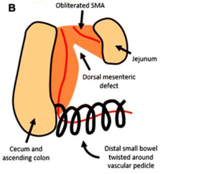
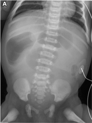
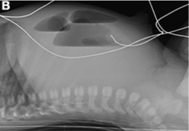
- Abdominal radiographs typically show a triple bubble appearance of only a few dilated loops of bowel within the upper abdomen, with air-fluid levels seen on the cross-table lateral view
Ileal atresia
- Ileal atresia occurs due to an ischemic insult in utero.
- Neonates typically present early in the neonatal period with vomiting, abdominal distention, and failure to pass meconium.
- Management is with surgical repair.
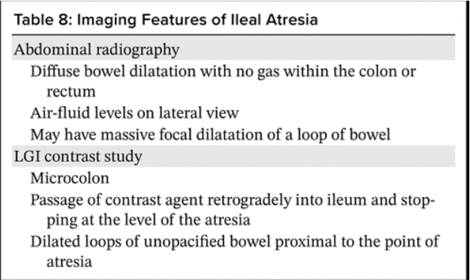
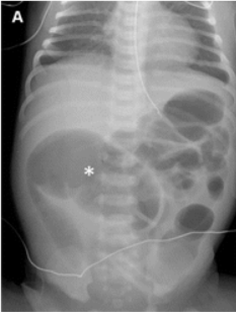
Diffuse dilatation bowel loops, with a more dilated loop in the right mid abdomen No gas is seen within the rectum
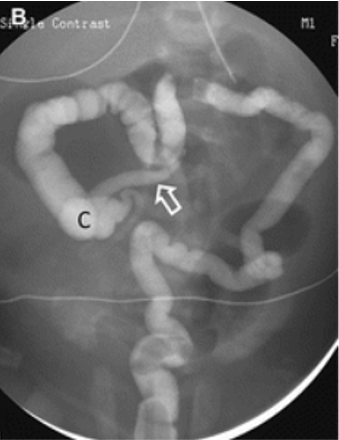
Microcolon with contrast material passing retrograde to the level of the cecum and into the distal ileum
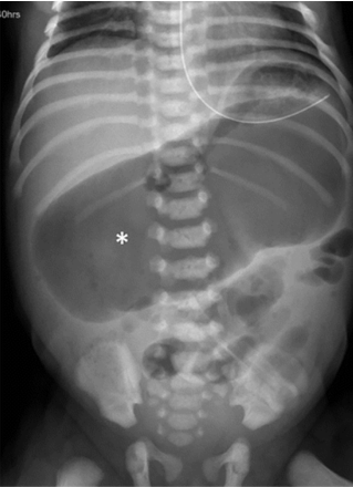
Massive focal dilatation of a loop of bowel within the mid and upper abdomen suggestive of obstruction secondary to atresia.
Differential diagnosis
- Functional immaturity of the large bowel.
- Meconium ileus
References:

Dr MADHU KUMAR SB
Senior consultant Radiologist
Manipal Hospital, Yeshwanthpur, Bengaluru.
Dr SHIKHA JOSHI
Radiology resident
Manipal Hospital, Yeshwanthpur, Bengaluru.

