
A 23 year old male presented with cough and dyspnoea since 3 months.
- A 23 year old male presented with cough and dyspnoea since 3 months.
- HRCT thorax was performed.
FINDINGS
- Numerous patchy nodular parenchymal infiltrates distributed diffusely in all the lung lobes, with a peribronchovascular and subpleural distribution.
- Apical emphysematous changes.
- No significant mediastinal lymphadenopathy seen. No effusion
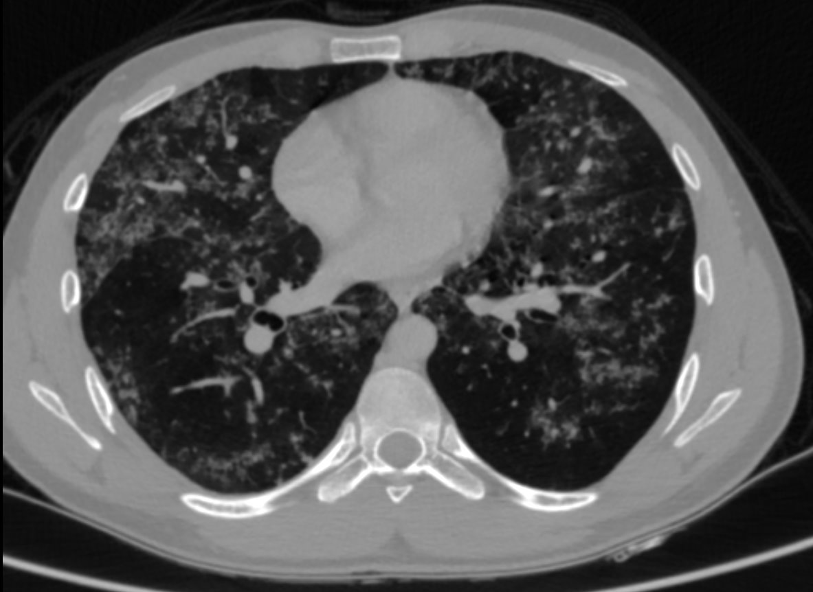
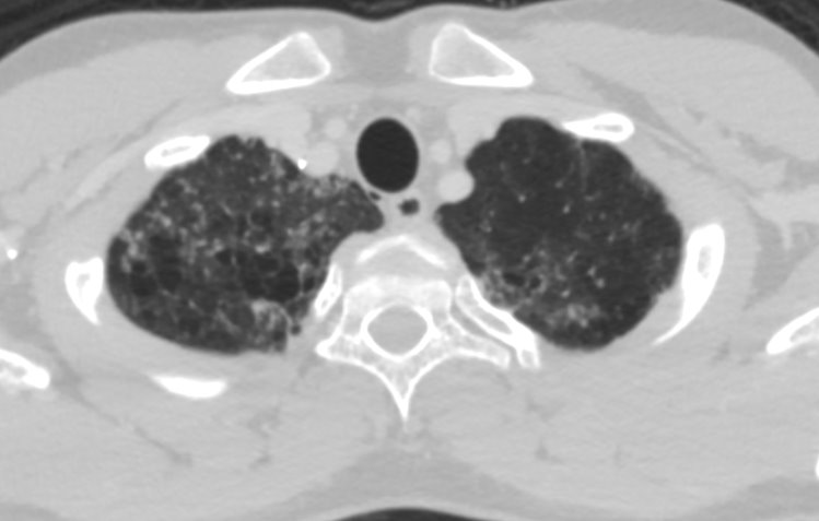
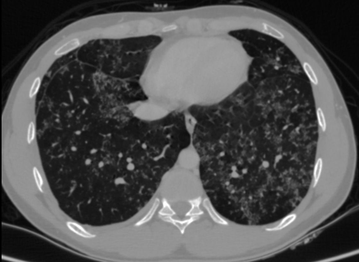
- Alveolar sarcoidosis
Alveolar sarcoidosis is an atypical pulmonary manifestation of sarcoidosis.
This appearance is thought to result from the aggregation of a vast number of interstitial granulomas rather than representing a true alveolar process.
Imaging Findings:
CT Features:
1.Appearance of alveolar sarcoidosis opacities:
- Patchy lung opacities 1–4 cm, rounded or elongated
- Irregular, blurred margins, may show air bronchograms
- Located along bronchovascular bundles or subpleural
2. Characteristic signs
- Galaxy sign: opacity with many surrounding small nodules (granulomas)
- Fairy ring / Reverse halo: circular arrangement of opacities with central clearing
3. Associated CT findings
- Nodules, ground-glass opacities,
- Thickened bronchovascular bundles,
- Thickened interlobular septa
References
-
Criado E, Sánchez M, Ramírez J, et al.
Pulmonary sarcoidosis: typical and atypical manifestations at high-resolution CT.
Radiographics. 2010;30(6):1567–1586.
(Classic paper describing galaxy sign, alveolar opacities, perilymphatic nodules) -
Hansell DM, Bankier AA, MacMahon H, et al.
Fleischner Society: glossary of terms for thoracic imaging.
Radiology. 2008;246(3):697–722.
(Defines reverse halo sign, ground-glass, opacities) -
Judson MA.
The diagnosis of sarcoidosis.
Clin Chest Med. 2008;29(3):415–427.
(General imaging findings in pulmonary sarcoidosis)
Dr Deepti HV
Senior Consultant Radiologist
Manipal Hospital, Yeshwantpur
Dr Nilay Gupta
Cross- sectional fellow
Manipal hospital , Yeshwantpur
