H/O SEIZURES.FIRST SEIZURES 15 YRS BACK.HEADACHE SINCE 15 DAYS.
HISTORY
- H/O SEIZURES.
- FIRST SEIZURES 15 YRS BACK.
- HEADACHE SINCE 15 DAYS.
FINDINGS -
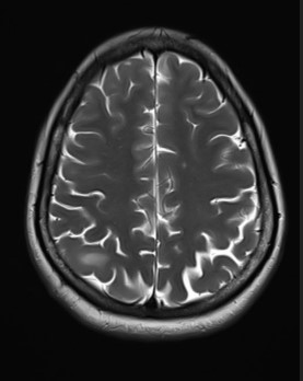
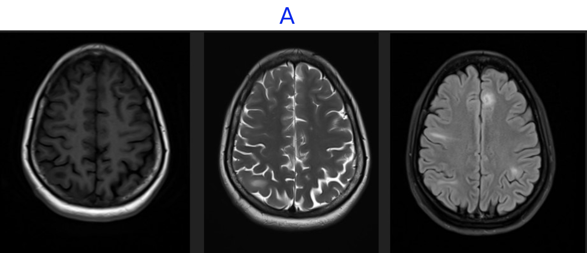
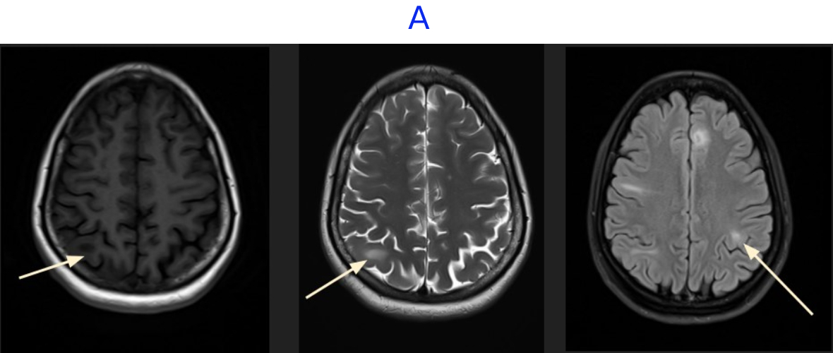
- A. MR BRAIN
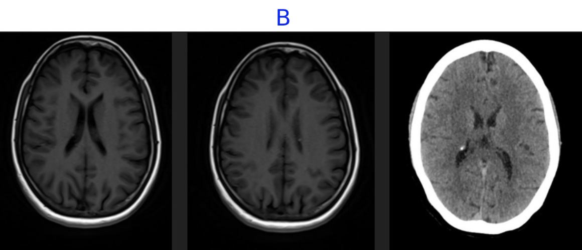
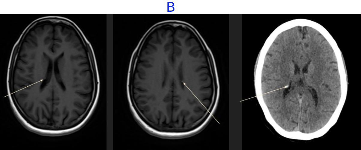
- B. MR AND CT BRAIN
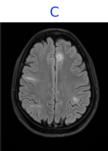
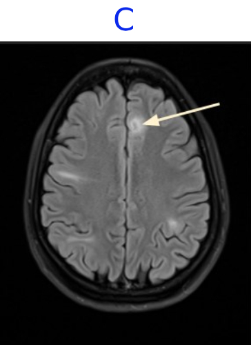
- C. MR BRAIN
LEGENDS
- A. There are multiple cortical and subcortical lesions seen in both cerebral hemispheres, most prominently involving the frontal and parietal lobes. These lesions demonstrate T2/FLAIR hyperintensity with corresponding T1 hypointensity, without associated mass effect. No evidence of any diffusion restriction or post-contrast enhancement.
- B. Multiple small subependymal nodules are seen along the margins of the bilateral lateral ventricles with few showing calcification on correlative CT.
- C. Areas of mild cortical thickening and blurring of the gray–white junction, consistent with focal cortical dysplasia
DIAGNOSIS
TUBEROUS SCLEROSIS
DISCUSSION
Tuberous sclerosis (TS), also known as tuberous sclerosis complex (TSC) or Bourneville disease, is a phakomatosis (neurocutaneous disorder) characterized by the development of multiple benign tumors of the embryonic ectoderm (e.g. skin, eyes, and central nervous system).
CLINICAL PRESENTATION
Tuberous sclerosis was classically described as presenting in childhood with a pathognomonic triad (Vogt triad) of:
- seizures: absent in one-quarter of individuals
- intellectual disability: up to half have normal intelligence
- adenoma sebaceum: only present in about three-quarters of patients
PATHOLOGY
Spontaneous mutations account for 50-86% of cases , with the remainder inherited as an autosomal dominant condition. In the majority of such cases (80%) the mutation has been narrowed down to two tumor suppressor genes, both part of the mTOR pathway 3,13:
- TSC1: encoding hamartin, on chromosome 9q32-34
- TSC2: encoding tuberin, on chromosome 16p13.3 (accounts for most
- cases)
RADIOLOGICAL FEATURES
Tuberous sclerosis has a significant number of manifestations, involving many organ systems. The most common radiographic manifestations are:
- cortical or subependymal tubers and white matter abnormalities
- renal angiomyolipomas
- cardiac rhabdomyomas
KEY FEATURES
CNS
- Cortical tubers
- SEN (calcified)
- SEGA (growing, enhances)
- Radial WM band
Renal
- AML (multiple, bilateral, hemorrhage risk)
- Cysts +/- early RCC
Thorax
- LAM (lung cysts, pneumothorax)
- Cardiac rhabdomyomas (infants; multiple = diagnostic)
Skin (95%)
- Ash-leaf spot
- Facial angiofibromas
- Shagreen patch
REFERENCES
- https://radiopaedia.org/articles/tuberous-sclerosis
- https://pubs.rsna.org/doi/abs/10.1148/rg.2021210103
DR. RAHUL KARTHIK LINGUTLA
CONSULTANT RADIOLOGIST,
MANIPAL HOSPITAL, YESHWANTHPUR
DR. FATHIMATH ASHILI KM
RADIOLOGY RESIDENT,
MANIPAL HOSPITAL, YESHWANTHPUR