A 57-year-old female came for follow-up for right buccal mucosa epidermoid carcinoma, post-surgery
A 57-year-old female came for follow-up for right buccal mucosa epidermoid carcinoma, post-surgery.
- 57 year old female.
- Follow up case of right buccal mucosa epidermoid carcinoma, post-surgery.

FINDINGS:
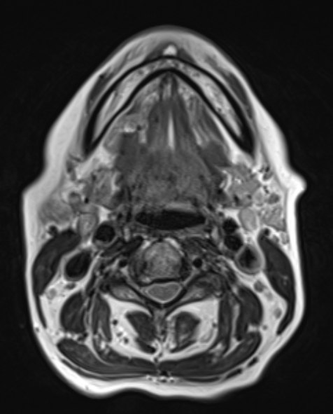
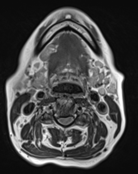
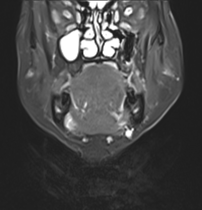
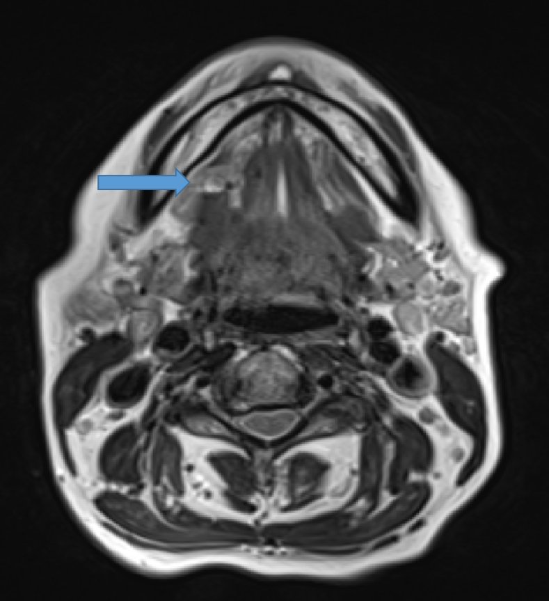
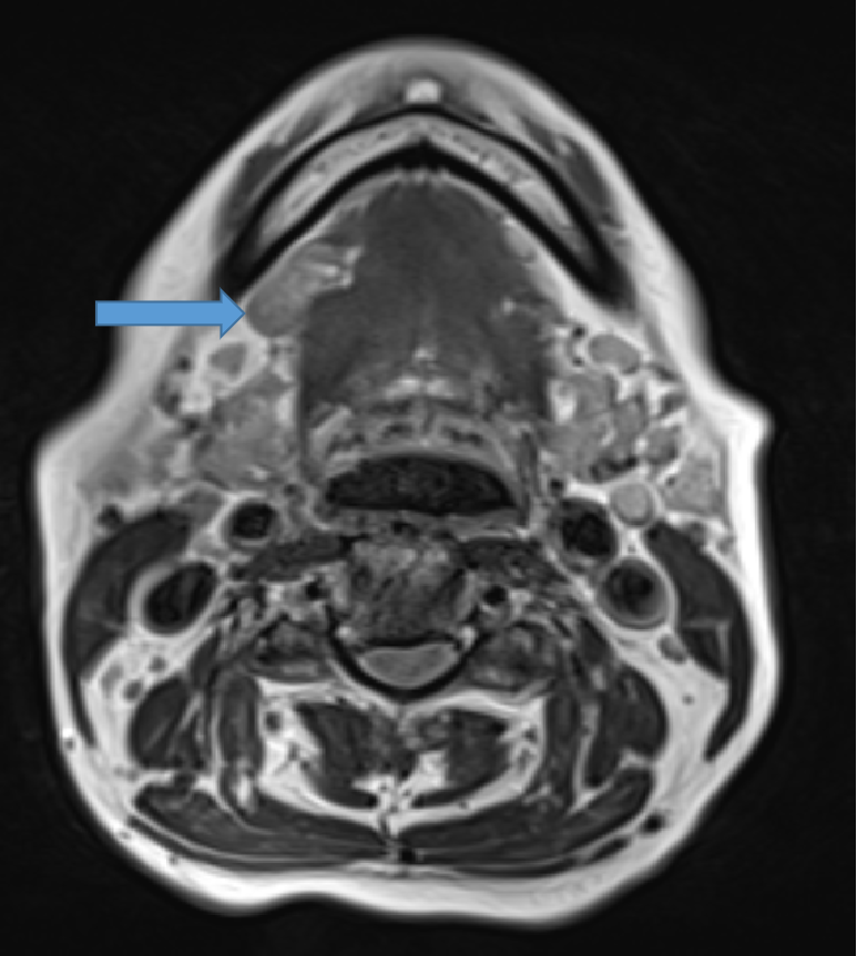
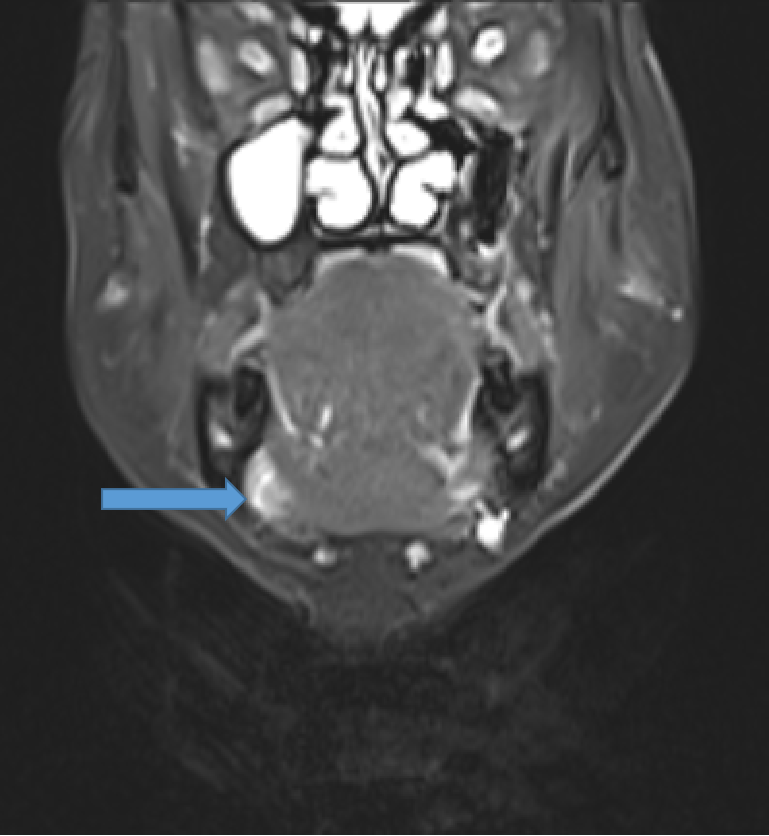
Coronal T2W images :
A. Defect noted in right side of mylohyoid muscle.
B. Herniation of right sublingual salivary gland from the mylohyoid muscle defect into submandibular space.
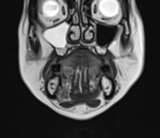
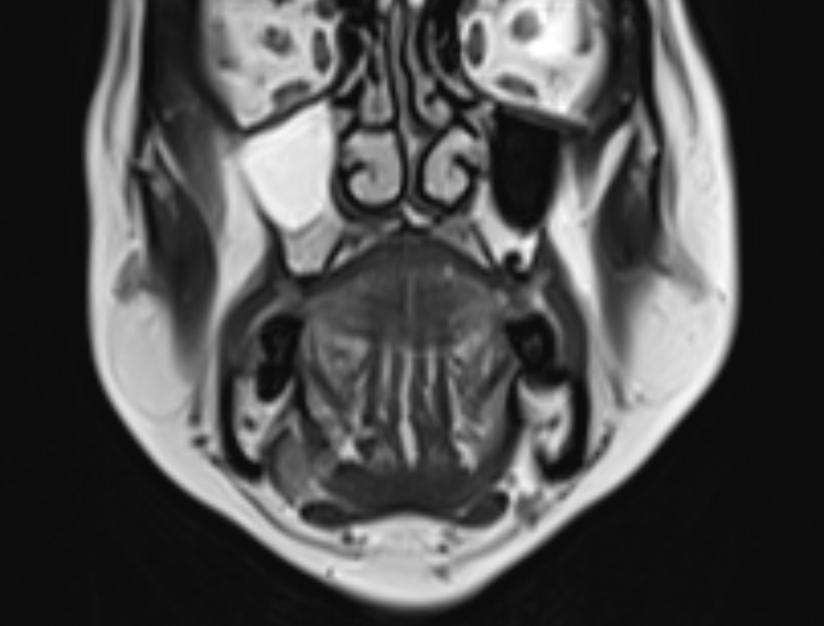
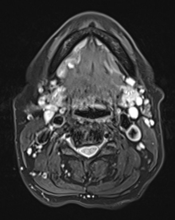
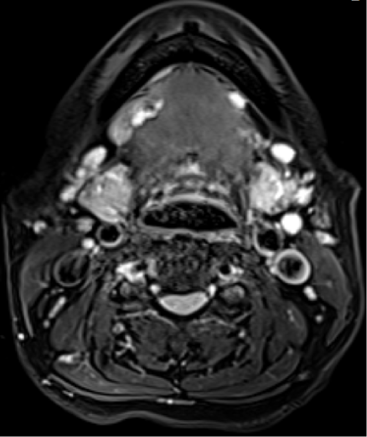
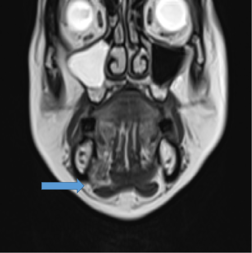
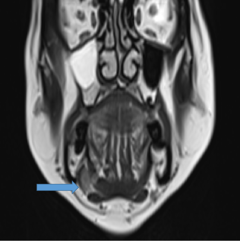
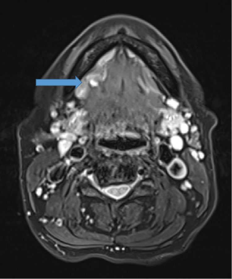
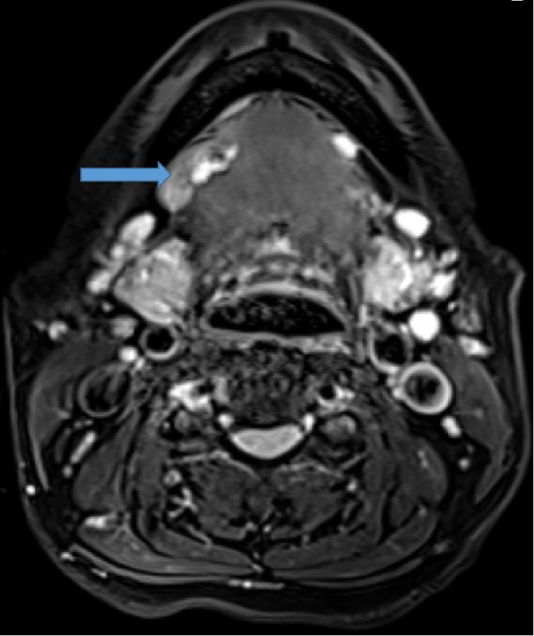
Axial T2W images:
A. Defect noted in right side of mylohyoid muscle.
B. Herniation of right sublingual salivary gland from the mylohyoid muscle defect into submandibular space.
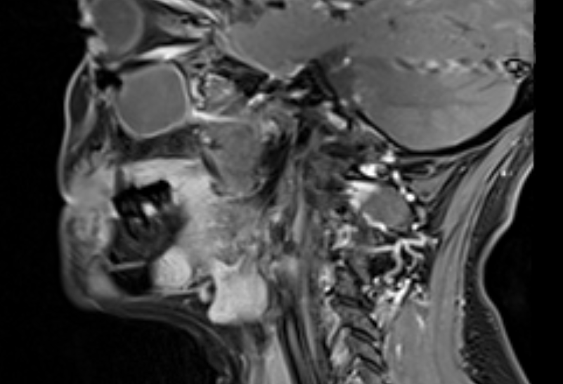
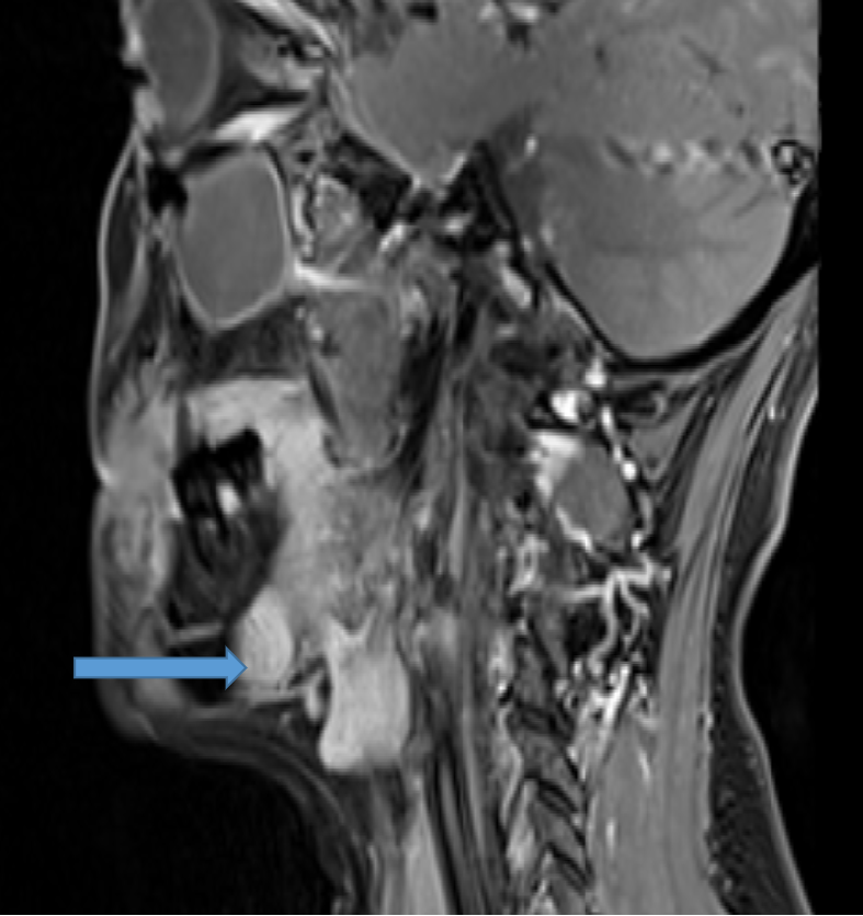
Axial and sagittal T2FSimages: Shows herniated right sublingual gland into sub mandibular space.
DIAGNOSIS:
- Mylohyoid boutonniere.
DISCUSSION:
- Mylohyoid boutonniere is a normal focal discontinuity in the Mylohyoid muscle, which may permit the sublingual salivary gland, fat or vessels - or a combination - to protrude out from the sublingual space into the submandibular space.
- The mylohyoid muscle is a sling-like structure that forms the floor of the oral cavity.
- This muscle divides the sublingual space from the sub mandibular space; however, communication between the two spaces posterior to the mylohyoid muscle is maintained.
- The mylohyoid muscle consists of two halves, both of which have a broad origin from the mylohyoid line along the inner mandible and the body of the hyoid bone.
- The two halves insert centrally at the fibrous median raphe to form a sling. Traditionally, the mylohyoid muscle is depicted as a continuous muscle sling.
- Most defects of the mylohyoid muscle are less than 5 mm, but occasionally they may be larger than 2 cm. Therefore, larger herniation may be mistaken both clinically and radiologically for pathologic abnormalities.
- Fat was the most common tissue identified within mylohyoid boutonnieres.
- Blood vessels were commonly identified traversing the mylohyoid muscle, both within and without identifiable defects.
- Discontinuity in the mylohyoid muscle, allowing normal structures of the sublingual space to protrude through the defect.
REFERENCES:
- White DK, Davidson HC, Harnsberger HR, Haller J, Kamya A. Accessory salivary tissue in the mylohyoid boutonnière: a clinical and radiologic pseudolesion of the oral cavity. American journal of neuroradiology. 2001 Feb 1;22(2):406-12.
- Iwai T, Sugiyama S, Ishikawa S, Mitsudo K. Sublingual Gland Herniation Masquerading as Submandibular Lesion. Indian Journal of Surgery. 2023 Apr;85(2):438-9.
- Sher ZA, Tan G. Unilateral sublingual salivary gland hypertrophy with herniation through a boutonniére defect and contralateral sublingual gland hypoplasia. BJR| case reports. 2016 Jul 28:20150382.
Dr. HARSHA CHADAGA
SENIOR CONSULTANT AND HEAD OF RADIOLOGY.
MANIPAL HOSPITAL,YESHWANTHPUR,BENGALURU
Dr. NIKITA PATEL
FELLOW IN CROSS SECTIONAL IMAGING
MANIPAL HOSPITAL,YESHWANTHPUR,BENGALURU