Frozen shoulder, medically known as adhesive capsulitis, is a notoriously painful condition that leads to progressive stiffness and agonizing loss of shoulder mobility.
While the core diagnosis is primarily established through clinical examination, radiology plays a massive role in ruling out underlying pathology, confirming the diagnosis and providing targeted interventional treatments.
Here is a breakdown of what happens inside the joint capsule, how it manifests across different modalities, and how interventional radiology is changing the treatment landscape.
The Pathology:
Frozen shoulder is characterized by inflammatory contracture of the shoulder joint capsule, leading to restriction of both active and passive ranges of motion.
The condition progresses through three different clinical phases:
| Phase | Timeline | Primary Pathological Event | Clinical Presentation |
|---|---|---|---|
| Freezing | 3 to 9 months | Acute synovitis & capsular inflammation | Severe, progressive pain; minor movement limits |
| Frozen | 4 to 12 months | Progressive fibrosis & capsular contracture | Pain plateaus; profound loss of active & passive ROM |
| Thawing | 12 to 42 months | Gradual resolution of fibrosis | Spontaneous, slow return of shoulder mobility |
What the Radiologist Sees: Imaging Findings
MRI is the gold standard imaging modality to diagnose this condition:
1. Magnetic Resonance Imaging (MRI)
MRI shows direct structural changes of adhesive capsulitis. The most reliable diagnostic keys include:
- Rotator Interval Obliteration: Infiltration and obliteration of the normal subcoracoid fat triangle inflammatory scar tissue or intense gadolinium enhancement.
- Axillary Pouch Thickening: The inferior glenohumeral ligament (IGHL) and axillary recess capsule show clear edematous thickening (typically >3 to 4 mm).
- Coracohumeral Ligament (CHL) Thickening: The CHL becomes distinctly thickened and indistinct.
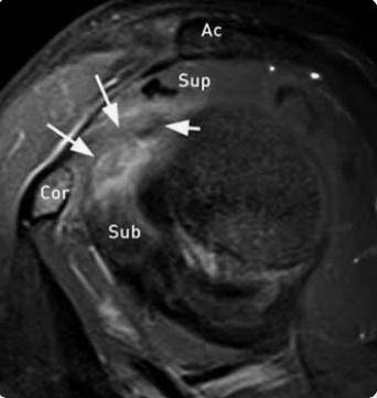
Soft tissue in Rotator interval
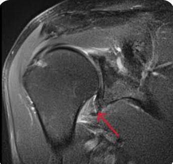
Edematous thickening of IGHL
Ultrasound (US)
High-resolution ultrasound provides a dynamic assessment. Radiologists look for a thickened coracohumeral ligament and increased vascularity within the rotator interval via power Doppler.
Interventional Radiology:
When conservative strategies like oral NSAIDs and physical therapy yield poor results, Interventional Radiology (IR) offers highly effective, image-guided alternatives that cut down recovery timelines significantly:
Hydrodissection (also known as hydrodilatation or high volume injection) is a highly effective, minimally invasive procedure used to treat frozen shoulder (adhesive capsulitis). The procedure involves injecting a controlled volume of sterile fluid into the shoulder joint under USG guidance to physically stretch the tight joint space, break down scar tissue and reduce inflammation.
Overview of the Procedure
| Attribute | Details |
|---|---|
| Primary Goal | Stretch the stiff joint capsule from within, rupture internal adhesions and restore range of motion. |
| Imaging Used | Ultrasound to ensure precision and safety during needle placement. |
| Typical Fluid Mix | A combination of sterile saline, a local anaesthetic (like lignocaine), and a corticosteroid (to suppress inflammation). |
| Fluid Volume | Typically 30 to 40 mL is introduced. A frozen shoulder joint's capacity shrinks to about 7 mL, so the fluid builds pressure until the capsule stretches or ruptures. |
| Success Rate | Provides significant pain relief in approximately 90% of patients and improves mobility in up to 70% of cases. |
| Duration & Setting | A 10 minute OPD procedure performed under local anaesthesia. Patient is observed for 15-20 mins after the procedure. |
What to Expect During the Procedure
The procedure is performed on an outpatient basis and typically follows these steps:
- Preparation: Patient will lie down, and the radiologist will clean the shoulder skin before applying a local anaesthetic to numb the needle track.
- Guidance: The radiologist uses real-time ultrasound to safely guide a thin spinal needle directly into the shoulder joint space.
- Distension: The fluid mixture is slowly injected. You may feel a deep, heavy pressure or tightness in the shoulder as the capsule expands. The procedure is complete once the desired volume is reached or the capsule gently ruptures, instantly relieving the internal pressure.
- Rehabilitation: Immediate, gentle physical therapy is strongly encouraged within days of the procedure to stretch the newly loosened joint and maintain the gained range of motion.
Advantages of Hydro dissection over traditional interventions
- Faster Recovery: Provides earlier, more rapid improvements in shoulder disability and sustained passive external rotation compared to standard corticosteroid injections alone.
- Less Invasive: Offers a much safer and cheaper alternative to aggressive surgical options like Manipulation Under Anaesthesia (MUA) or arthroscopic capsular release.
- Immediate Relief: Many patients note an immediate reduction in mechanical stiffness right after the fluid expands the joint space
Important Considerations for Diabetics
Individuals with diabetes have a significantly higher incidence of frozen shoulder (up to 20%). Corticosteroid used in the solution may cause a transient spike in blood glucose levels. Diabetics must closely monitor their blood sugar levels for at least 48 hours to a week following the injection and may coordinate with physician regarding temporary insulin or medication adjustments.