


A Radiologist’s Perspective
Not every disc bulge causes pain, and not every patient with severe back pain has a significant abnormality on MRI. The key lies in correlating imaging findings with clinical symptoms.
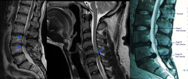
Back pain, neck pain and radiating limb pain are common reasons for medical consultation. Although many cases improve with conservative management, persistent symptoms or neurological manifestations may warrant further investigation. Magnetic Resonance Imaging (MRI) plays an important role in evaluating spinal disc pathology by providing detailed anatomical information about the intervertebral discs, spinal canal, neural foramina, nerve roots and spinal cord.
From a radiologist’s perspective, the value of MRI extends beyond identifying structural abnormalities. Its clinical relevance lies in determining whether the observed findings correspond to the patient’s symptoms and provide useful information for diagnosis and treatment planning.
1. Understanding Spinal Disc Pathology
Intervertebral discs are specialised structures situated between adjacent vertebral bodies. They provide load distribution, shock absorption and flexibility to the spine. With ageing and degeneration, these discs may undergo changes in hydration, structure and mechanical properties. Trauma and other factors may also contribute to disc pathology.
MRI helps characterise several important abnormalities:
- Disc desiccation: Reduced water content within the disc, typically demonstrated by decreased T2-weighted signal intensity.
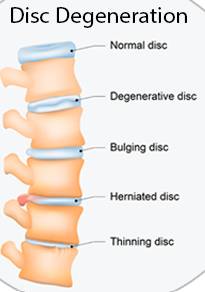
- Disc degeneration: Structural changes that may include altered disc signal, loss of disc height and associated endplate abnormalities.
- Disc bulge: Generalised extension of disc tissue beyond the vertebral body margins.
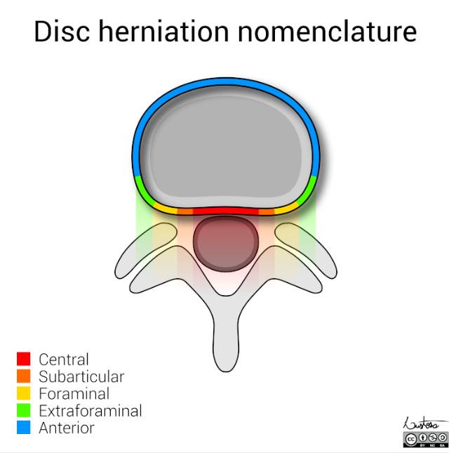
- Disc protrusion: A form of localised disc herniation in which the displaced disc material has a relatively broad base compared with its outward extension.
- Disc extrusion: Herniated disc material extending beyond the disc space with a narrower connection to its origin or other defining morphological features.
- Annular fissure: A disruption or separation within the annular fibres.
- Spinal canal or neural foraminal stenosis: Narrowing that may compromise the spinal cord or nerve roots, depending on its location.
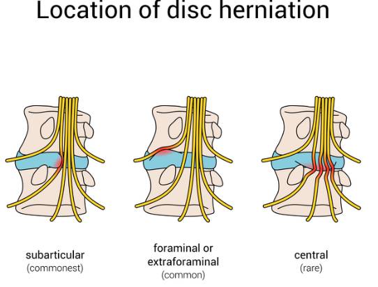
These findings must be interpreted in the context of their location, extent and relationship to adjacent neural structures.
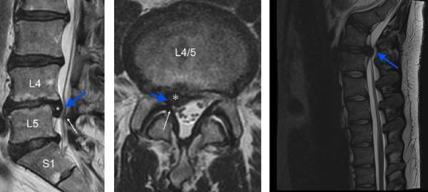
2. The Radiologist’s Challenge: Establishing Clinical Relevance
Consider two patients with lumbar disc abnormalities.
The first patient has a disc bulge on MRI but no corresponding radicular symptoms or neurological deficits. The second patient presents with lower back pain radiating into the leg, accompanied by paraesthesia or weakness. MRI demonstrates a disc herniation that compresses a nerve root corresponding to the clinical distribution of symptoms.
Although both patients have disc pathology, the clinical significance of their findings may differ substantially. This illustrates why MRI interpretation must go beyond simply identifying a disc bulge.
A systematic radiological assessment includes the affected spinal level, morphology and location of the disc abnormality, degree of neural compromise, associated degenerative changes and any additional findings that may influence clinical management. The objective is to determine whether the imaging findings provide a plausible anatomical explanation for the patient’s symptoms, rather than assuming that every abnormality is symptomatic.
3. How MRI Helps Evaluate Back Pain and Radicular Symptoms
A. Disc degeneration and axial back pain
Degenerative changes involving the intervertebral discs and adjacent vertebral endplates may be associated with axial back pain in selected patients.
However, similar changes are frequently observed in asymptomatic individuals. Therefore, the presence of disc degeneration alone does not establish the source of pain. Clinical correlation remains essential.
B. Disc herniation and radiculopathy
A herniated disc may cause radicular symptoms through mechanical compression, inflammatory irritation or a combination of both.
Lumbar nerve root involvement may produce sciatica, with pain radiating from the lower back into the buttock and leg. Cervical nerve root involvement may cause pain, paraesthesia or altered sensation extending into the shoulder and upper limb.
MRI helps identify the level and location of the herniation and assess its relationship to the corresponding nerve root. These findings can support clinical diagnosis when they correlate with the distribution of symptoms and neurological examination.
C. Spinal stenosis and neural compression
Degenerative disc changes may coexist with facet joint arthropathy, osteophyte formation and ligamentum flavum thickening. Together, these abnormalities may contribute to central canal, lateral recess or neural foraminal stenosis.
MRI enables assessment of the location and extent of narrowing and helps determine whether the spinal cord or nerve roots are affected. In patients with cervical spinal stenosis, particular attention should be paid to spinal cord compression and associated cord signal abnormalities, which may indicate myelopathy.
4. How MRI Contributes to Treatment Planning
MRI findings can support treatment decisions when interpreted alongside the clinical history, physical examination and functional limitations.
A. Conservative management
Many patients with uncomplicated back pain or disc herniation can initially be managed without surgery. Depending on the clinical presentation, treatment may include appropriate physical activity, structured rehabilitation, physiotherapy and clinician-directed pain management.
MRI is not routinely required for every episode of uncomplicated acute back pain. It becomes particularly useful when persistent symptoms, neurological findings or suspected serious pathology make imaging clinically appropriate.
B. Image-guided pain interventions
In selected patients with persistent radicular pain, MRI may help identify the suspected symptomatic level before an epidural steroid injection or selective nerve root injection is considered. Detailed anatomical information can assist procedural planning and help clinicians assess whether the proposed intervention is appropriate. However, imaging findings alone cannot reliably predict treatment response, and the decision to intervene must be based on the overall clinical assessment.
C. Surgical planning
Surgical evaluation may be appropriate when symptoms remain disabling despite suitable conservative treatment or when significant neurological compromise is present.
MRI provides important information regarding:
- The level, morphology and extent of disc herniation.
- The location and degree of nerve root compression.
- The severity and distribution of spinal canal or foraminal stenosis.
- Associated degenerative changes.
- Spinal cord compression and relevant cord signal abnormalities.
These findings help surgeons assess the anatomical basis of symptoms and plan appropriate procedures in selected patients. An MRI abnormality alone is not an indication for surgery. The decision must be guided by clinical findings, symptom severity, functional impairment and the expected benefits and risks of intervention.
5. Can MRI Predict the Severity of Pain?
One of the important limitations of spinal MRI is that the severity of imaging abnormalities does not always correlate with the severity of symptoms. Some individuals demonstrate substantial degenerative changes without experiencing significant pain. Conversely, patients with considerable pain may have relatively subtle structural abnormalities on MRI.
Pain is multifactorial and may involve mechanical, inflammatory, neurological, biological and psychosocial influences that cannot be fully characterised by conventional imaging. Consequently, MRI should not be regarded as an independent measure of pain severity. Its greatest diagnostic value emerges when imaging findings are integrated with clinical history, neurological examination and the patient’s functional status.
6. The Importance of Structured Radiological Reporting
A clinically useful MRI report should do more than list degenerative changes. It should clearly communicate the location and morphology of relevant abnormalities, their relationship to neural structures and the presence of associated findings that may influence management.
For example, describing a disc herniation at a specific level and documenting its relationship to the traversing or exiting nerve root may provide more useful clinical information than simply reporting a disc bulge. Similarly, distinguishing incidental degenerative changes from findings that plausibly explain the patient’s symptoms help reduce the risk of unnecessary concern or inappropriate intervention. When appropriate, the report should also highlight significant spinal cord compression, suspected infection, tumour or other potentially urgent pathology.
7. The Role of Radiology in Multidisciplinary Patient Care
Effective management of spinal disc pathology often requires collaboration between radiologists, referring clinicians, physiotherapists, pain specialists and spinal surgeons. The radiologist contributes detailed anatomical assessment, identifies potentially significant abnormalities and communicates findings relevant to diagnosis and treatment planning.
The treating clinician integrates this information with the patient's symptoms, examination findings, functional limitations and response to previous treatment. This multidisciplinary approach helps ensure that imaging supports patient-centred decision-making rather than becoming the sole determinant of treatment.
Conclusion: From Imaging Findings to Clinical Decisions
Spinal MRI is an essential diagnostic tool for evaluating disc pathology, neural compression and degenerative spinal disorders when clinically indicated. However, its true value lies not simply in identifying abnormalities, but in establishing their potential clinical significance.
A disc bulge is an imaging finding. Nerve root compression is an anatomical observation. Determining whether either finding explains a patient’s symptoms require careful clinical correlation.
From a radiologist’s perspective, the ultimate goal is to provide accurate, structured and clinically relevant information that helps guide appropriate treatment, avoid unnecessary interventions and improve patient care.
The objective of spinal imaging is not merely to identify pathology, but to translate anatomical information into meaningful clinical decisions.