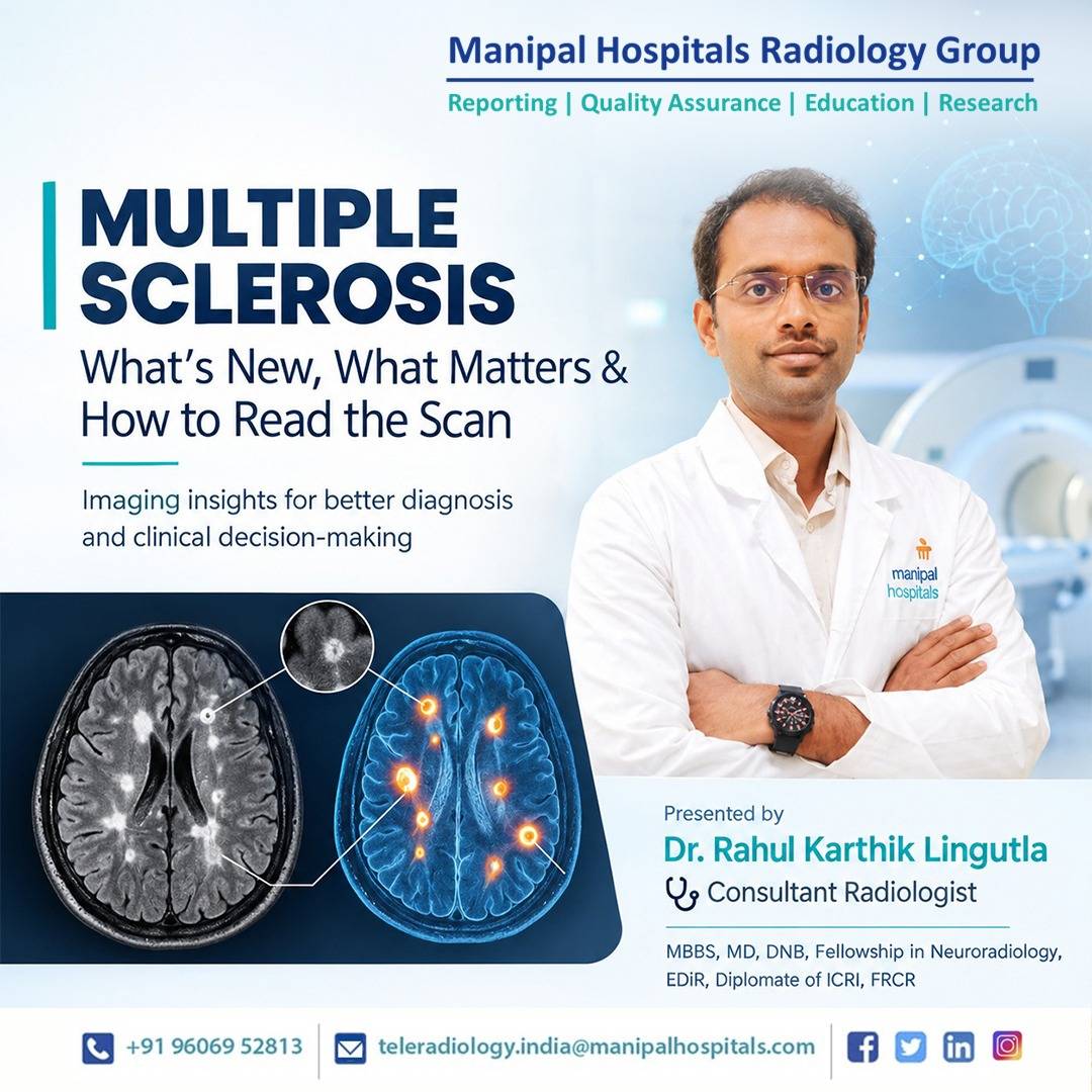
Multiple sclerosis (MS) remains one of the most important inflammatory demyelinating diseases in neuroradiology. For radiologists, MS is no longer just about spotting “typical white matter lesions.” With the 2024 revision of the McDonald criteria, advanced MRI biomarkers, and a more nuanced approach to radiologically isolated syndrome (RIS), your report can directly influence diagnosis, treatment timing, and even whether a patient is labeled as having MS at all.
This post walks through what you need to know to read MS studies with confidence in 2026: updated diagnostic criteria, key MRI patterns, high-specificity signs, pitfalls, and practical reporting tips.
Why the 2024 McDonald Criteria Matters:
The McDonald criteria are the backbone of MS diagnosis, integrating clinical, imaging, and laboratory data. The 2024 update introduces several changes that elevate the role of MRI and specific biomarkers.
Key updates:
- Optic nerve added as a fifth topographic site for dissemination in space (DIS), alongside periventricular, juxtacortical/cortical, infratentorial, and spinal cord regions.
- Stricter thresholds in older patients and those with vascular risk to reduce misdiagnosis, particularly in patients >50 years or with headache/vascular comorbidities.
- Radiologically isolated syndrome (RIS) can now be classified as MS under specific conditions, shifting some cases from “incidental finding” to a treatable disease.
- Two high-specificity MRI biomarkers formally incorporated: the central vein sign (CVS) and paramagnetic rim lesions (PRLs) on susceptibility-weighted sequences.
- CSF kappa free light chains (kFLC) accepted as an alternative to oligoclonal bands (OCBs) for demonstrating intrathecal immunoglobulin synthesis.
In practice, this means your description of lesion location, morphology, and susceptibility characteristics now carries more diagnostic weight than before.
Recognizing Typical MS Lesions: Location and Morphology
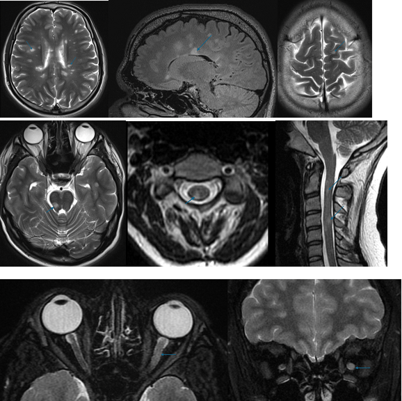
MS lesions are classically:
- Ovoid, well-circumscribed T2/FLAIR hyperintensities
- Often perpendicular to the ventricles (“Dawson’s fingers”) in the periventricular white matter
- Frequently involving the corpus callosum(callosal-septal interface)
- Common in the juxtacortical/cortical region, including U-fibers
- Can involve the infratentorial compartment (brainstem, cerebellum, middle cerebellar peduncles)
- In the spinal cord, typically short-segment (<2 vertebral bodies), peripheral, and involving less than half the cross-sectional area.
Under the McDonald criteria, demonstrating dissemination in space (DIS) requires typical lesions in at least two of five regions (periventricular, juxtacortical/cortical, infratentorial, spinal cord, optic nerve).
High-Specificity MRI Biomarkers: CVS and Paramagnetic Rim Lesions
Central Vein Sign (CVS)
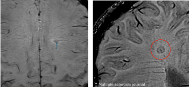
The CVS refers to a small central vein traversing a white matter lesion, best seen on SWI sequences. It reflects the perivenular inflammatory nature of MS lesions.
Key points:
- ≥6 CVS-positive lesions in the brain can serve as a stand-alone biomarker supporting MS diagnosis, including in RIS or non-specific presentations.
- If the total lesion count is <10, the majority of lesions should be CVS-positive to maintain specificity.
- CVS helps distinguish MS from mimics such as small-vessel ischemic disease, migraine-related changes, and some inflammatory or infectious leukoencephalopathies.
In your report, it can be helpful to explicitly state whether lesions demonstrate a central vein when this is clear, especially in diagnostically challenging cases.
Paramagnetic Rim Lesions (PRLs)
PRLs appear as T2-hyperintense lesions with a hypointense rim on SWI, corresponding to iron-laden macrophages at the lesion edge. They are associated with chronic active (“smoldering”) lesions and more aggressive disease.
- PRLs are now recognized as high-specificity markers integrated into the 2024 criteria.
- Their presence supports MS over mimics and may carry prognostic information regarding progression.
Noting the presence of PRLs in suspicious cases can add value, particularly when the clinical picture is atypical.
Radiologically Isolated Syndrome: When “Incidental” Becomes MS
RIS is defined by incidental MRI findings typical of MS in a patient without clinical demyelinating symptoms. Historically, RIS was a radiologic observation, not a diagnosis of MS. The 2024 criteria change that for a subset of patients.
Under the updated McDonald criteria, RIS can be diagnosed as MS when:
- DIS is fulfilled (typical lesions in at least two of five regions), and
At least one of the following is present:
- Dissemination in time (DIT) on MRI (new T2 lesion or gad-enhancing lesion compared to prior)
- Positive CSF (OCBs or elevated kFLC index)
- ≥6 CVS-positive lesions (or majority CVS+ if <10 total lesions)
This suggests:
- Careful characterization of lesion distribution and morphology is critical.
- Explicitly stating whether lesions are “typical of MS” and whether they fulfil DIS can directly impact whether the patient meets criteria for MS or remains labelled as RIS.
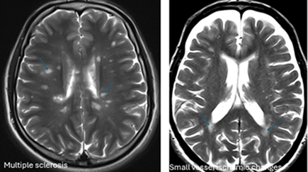
Differentiating MS Plaques from chronic small-Vessel Ischaemic and Other Non-Specific White Matter Changes
Residents and young radiologists often ask how to differentiate multiple sclerosis demyelinating plaques from small-vessel ischaemic changes and other non-specific white matter hyperintensities commonly seen in the cerebral white matter.
MS plaques typically occur in very typical locations: juxtacortical, periventricular, infratentorial, and spinal cord regions. Lesions in these typical locations favour a demyelinating aetiology rather than chronic small-vessel ischaemic change.
Particularly in the periventricular region, certain morphological features strongly support a demyelinating plaque:
- Orientation parallel to the ventricular margin, rather than the perpendicular, cap-like orientation often seen with Dawson’s fingers (note: classic Dawson’s fingers are perpendicular; many small-vessel changes are more confluent and cap-like along the ventricular surface)
- Involvement of the calloso-septal interface
- Triangular or wedge-shaped morphology of the T2/FLAIR hyperintensity extending toward the cortex
Looking at these typical locations and the morphology of the white matter hyperintensity will help to differentiate between a demyelinating plaque versus chronic small-vessel ischaemic change or other non-specific hyperintensities. In older patients or those with significant vascular risk factors, the 2024 McDonald criteria explicitly call for stricter thresholds before diagnosing MS, making these distinctions even more important.