
A 42-year-old man presented with complaints of right calf pain
A 42-year-old man presented with complaints of right calf pain
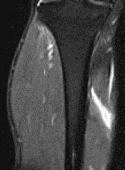
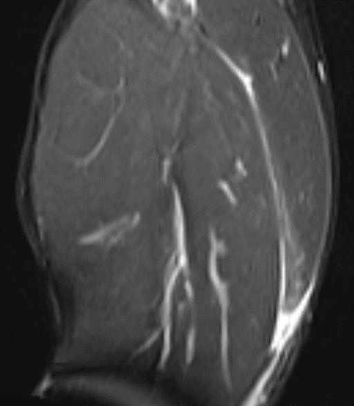
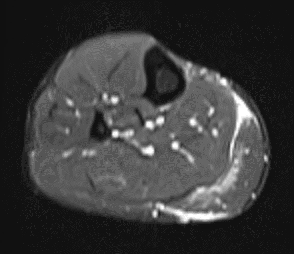
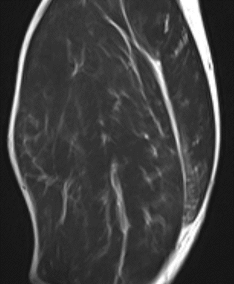
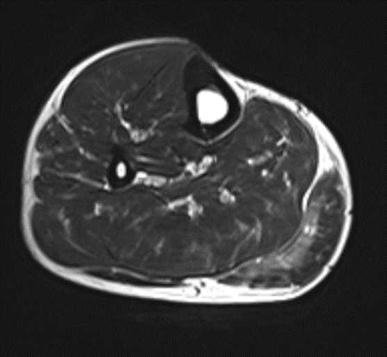
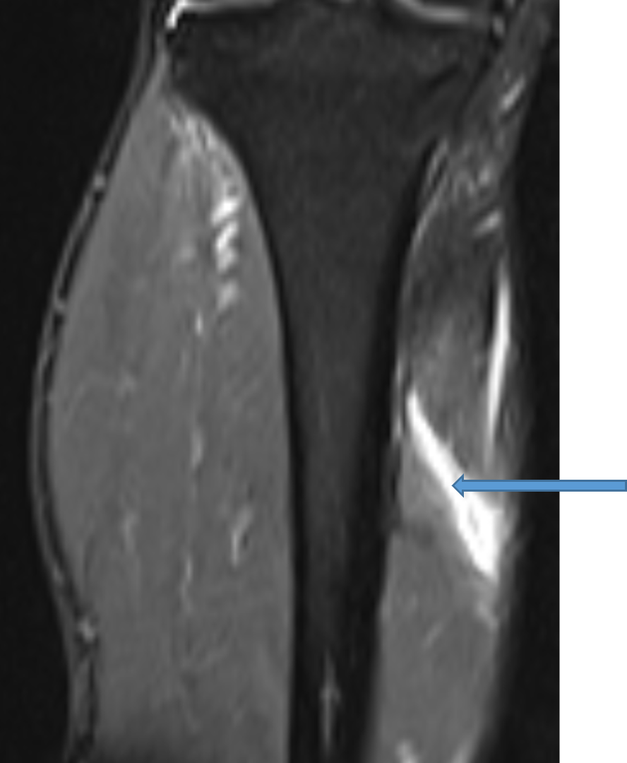
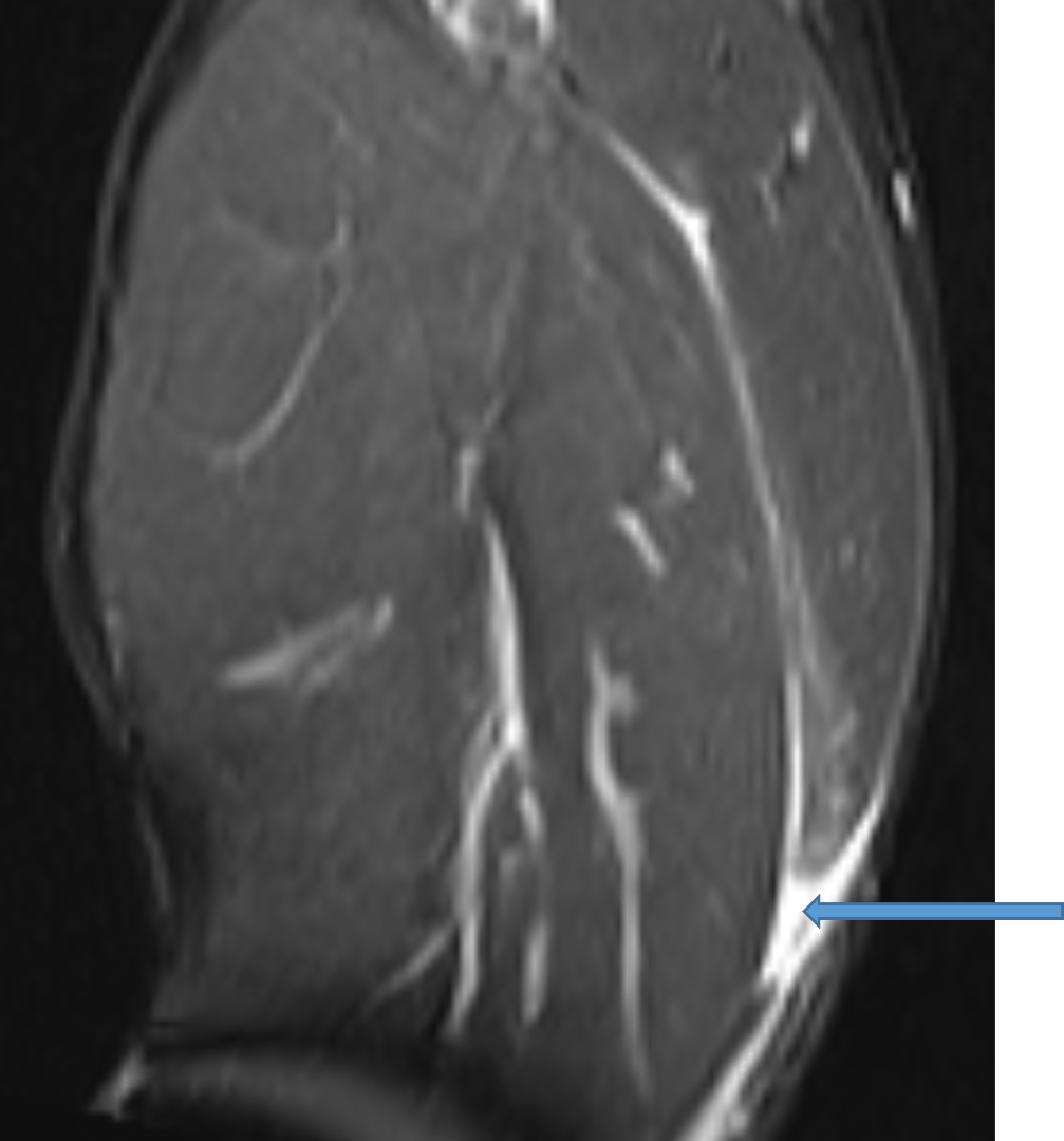
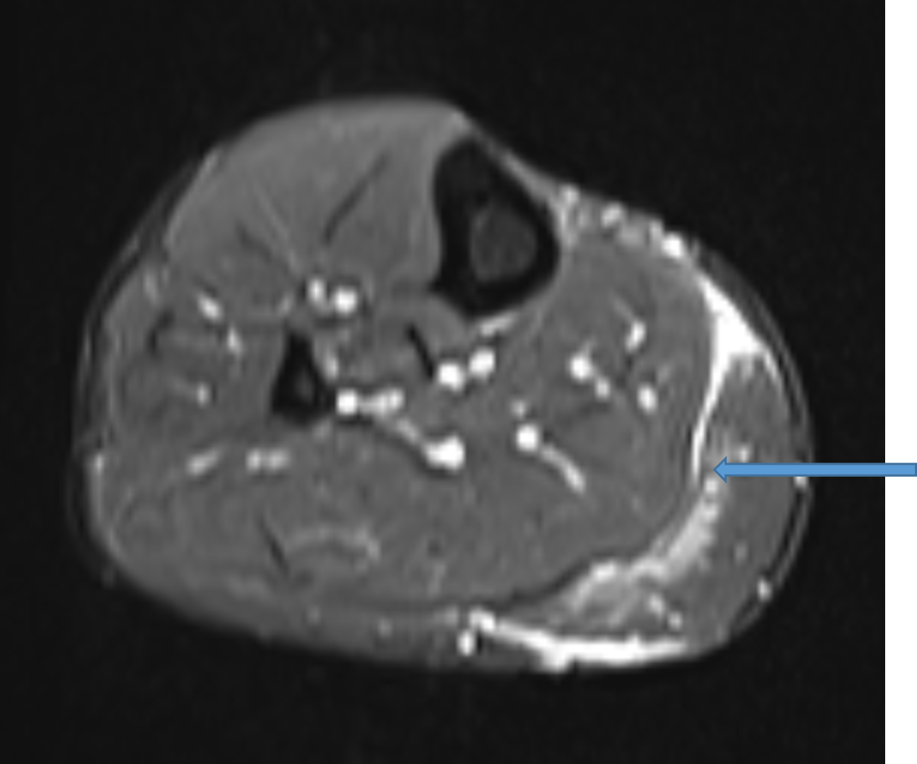
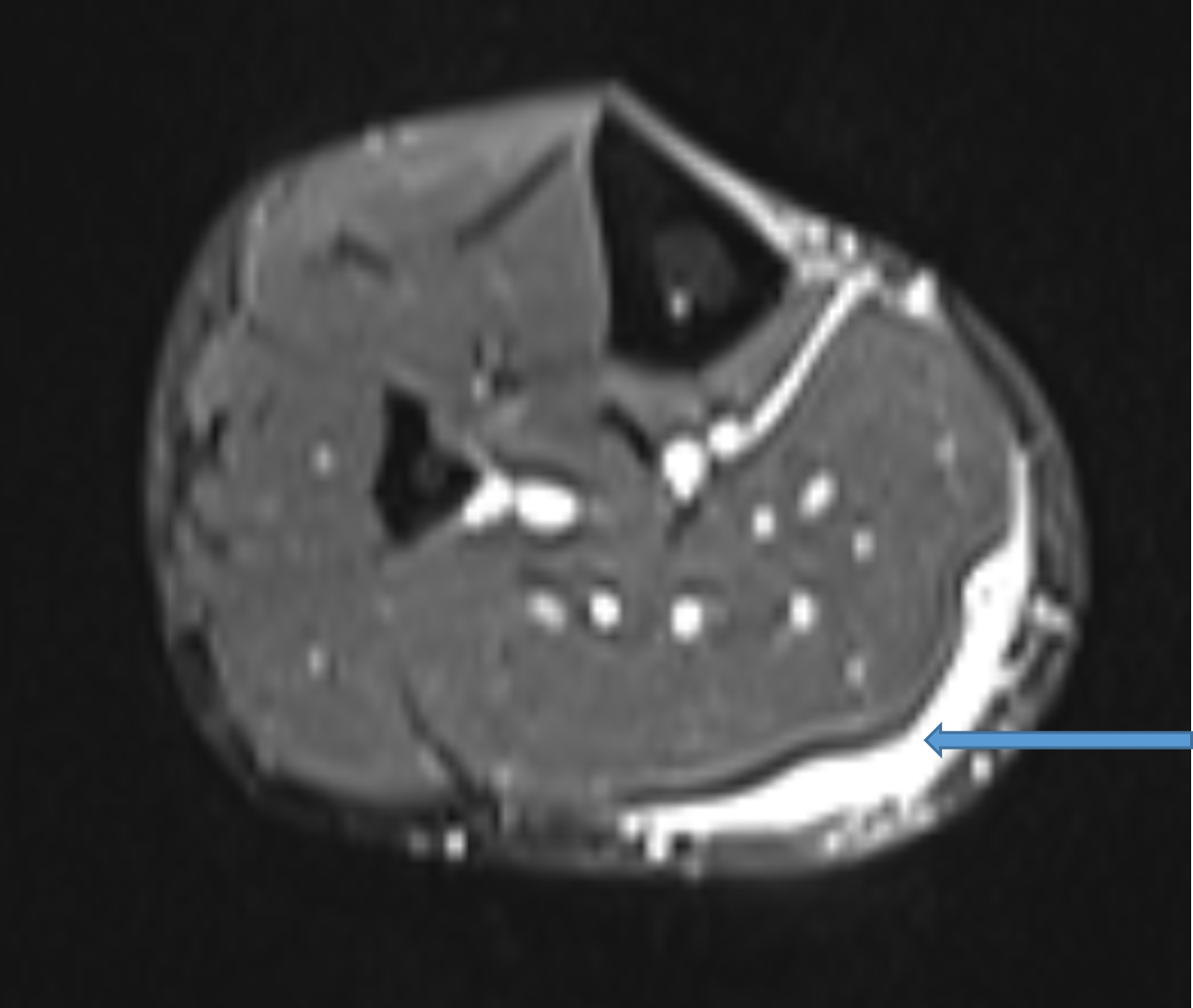
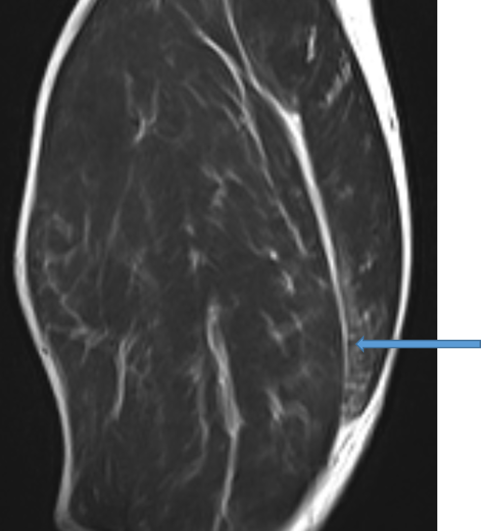
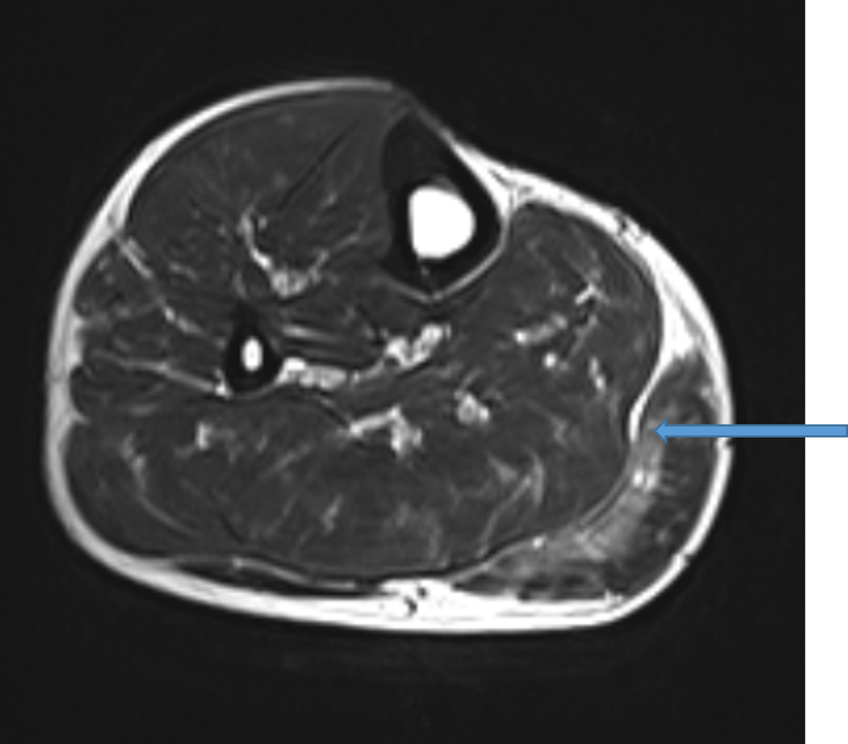
- STIR and T2 images showing hyper intensity with partial discontinuity in the deep fibers of the medial head of gastrocnemius muscle at the musculotendinous junction
- Minimal fluid collection is seen insinuated between the distal medial head of the gastrocnemius muscle and the soleus muscle with edema.
Diagnosis : Tennis Leg
DISCUSSION:
- Tennis leg represents a myo-fascial or tendinous injury of the lower limb .
- Seen most frequently in tennis players.
Pathology :
It involves injury to the muscles of the calf (superficial posterior compartment of the leg) via two identified mechanisms:
- Tear of the myotendinous junction of the medial head of the gastrocnemius
- Rupture of the plantaris tendon
Both findings have been found in isolation or together.
Imaging:
Radiographic features
Ultrasound
- Ultrasound demonstrates fluid deep to medial gastrocnemius and superficial to the soleus muscle.
- Most prominent at the level of the myotendinous junction.
- A tear in the deep surface of gastrocnemius may be seen as a disruption in contour and echogenicity of muscle fibers.
- A torn plantaris tendon may also be identified.
MRI
MRI demonstrates the same features seen on ultrasonography. Findings include:
- High T2 signal fluid deep to medial gastrocnemius and superficial to the soleus
- A focal area of disruption of muscle continuity noted along the deep aspect of the medial head of the gastrocnemius, with associated muscle edema
- The plantaris tendon may either be torn or intact
DIFFERENTIAL DIAGNOSIS
- The differential diagnosis of calf pain includes the more serious clinical entities of deep venous thrombosis (DVT) and compartment syndrome, as well as Achilles tendon rupture.
- In the proper clinical situation, calf pain with an otherwise negative MRI raises suspicion for DVT. An Achilles tendon rupture is easily differentiated from plantaris tendon rupture on MRI.
- Compartment syndrome can also be distinguished from plantaris or gastrocnemius muscle injury, since it demonstrates a more diffuse muscle edema pattern.
- Compartment syndrome, however, may be seen in conjunction with calf muscle injuries, as a result of hematoma and swelling
References :
- Tennis leg. Br Med J. 1969;3 (5670): 543-4. Free text at pubmed- Pubmed citation
- Delgado GJ, Chung CB, Lektrakul N et-al. Tennis leg: clinical US study of 141 patients and anatomic investigation of four cadavers with MR imaging and US. Radiology. 2002;224 (1): 112 9. doi:10.1148/radiol.2241011067 – Pubmed citation
- Jamadar DA, Jacobson JA, Theisen SE et-al. Sonography of the painful calf: differential considerations. AJR Am J Roentgenol. 2002;179 (3): 709-16. AJR Am J Roentgenol (full text) – Pubmed citation
- Bianchi S, Martinoli C, Abdelwahab IF et-al. Sonographic evaluation of tears of the gastrocnemius medial head (“tennis leg”) J Ultrasound Med. 1998;17 (3): 157-62. J Ultrasound Med (abstract) – Pubmed citation
- Shah JR, Shah BR, Shah AB. Pictorial essay: Ultrasonography in ‘tennis leg’. Indian J Radiol Imaging. 2010;20 (4): 269-73. doi:10.4103/0971-3026.73542 – Free text at pubmed- Pubmed citation
Dr. PRAVEEN WALI
Senior Consultant Radiologist
Manipal Hospital, Yeshwanthpur, Bengaluru.
Dr. Srinivas P
Fellow in Radiology
Manipal Hospital Radiology Group (MHRG)
Manipal Hospital, Bengaluru