
25 year old gentleman with history of fall, pain and swelling in right knee
25-year-old gentleman with history of fall, pain, and swelling in his right knee.
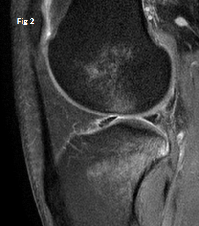
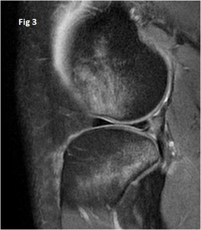
Figure 1: PDSF sagittal MR image demonstrates near complete tear of mid anterior cruciate ligament.
Figure 2 & 3: PDFS sagittal image demonstrates longitudinal fluid signal cleft at the periphery of posterior horn of lateral meniscus at the attachment of ligament of Wrisberg extending far laterally in next image. Pivot shift contusions also seen.
DIAGNOSIS:
Near complete tear of anterior cruciate ligament with Wrisberg rip.
Discussion:
- LIGAMENT OF WRISBERG – A meniscofemoral ligament coursing posterior to posterior cruciate ligament as it attaches to the posterior horn of lateral meniscus.
- This case demonstrates a particular lateral meniscal tear pattern occurring in association with ACL tears where a longitudinal tear is noted within the posterior horn of lateral meniscus extending laterally from Wrisberg ligament attachment site: “Wrisberg rip”
- Wrisberg pseudo-tear: Normal vertical fluid cleft seen at the junction of posterior horn of lateral meniscus and ligament of Wrisberg. This is a normal anatomical appearance and should not be called a tear.
Practical points:
- Fluid cleft that extends far lateral from the ligament attachment site on consecutive sagittal cuts: likely Wrisberg rip.
- Average attachment site of Wrisberg ligament lies approximately 14 mm laterally from the lateral edge of the PCL , and that any cleft extending farther than this point suggests Wrisberg rip.
Wrisberg rip must be carefully looked for in every case of ACL tear, on the other hand presence of Wrisberg rip serves as a secondary sign of ACL disruption.
Dr Dayananda Sagar, MD
Consultant Radiologist
Dr Shashwat Priyadarshi, DNB
Cross-sectional imaging fellow