
2 year old presenting with leucoria and proptosis
2 year old presenting with leucoria and proptosis
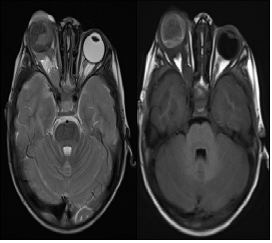
- Images 1: Axial T2 and T1 images demonstrate a T2/T1 hyperintense right intraocular mass with the large exophytic component, distorting the right globe with proptosis.
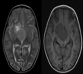
- Images 2: Axial T2 and T1 images demonstrate well-defined extra-axial mass lesions in the suprasellar region with internal cystic changes.
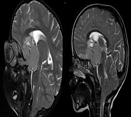
- Images 3: Oblique sagittal STIR and T2 sagittal images demonstrate the extra-scleral extension of the lesion along the right optic nerve involving the optic chiasma and contiguity with the supra-sellar lesion.
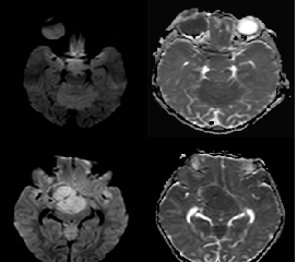
- Images 4: Axial SWI and ADC images demonstrate hyperintensity on DWI with the corresponding dark signal on ADC in the intraocular and suprasellar lesion.
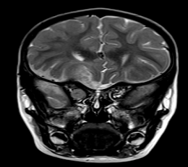
- Image 5: Coronal T2 image demonstrates patchy and leptomeningeal spread of lesion along the right frontal lobe
DIAGNOSIS:
Right ocular Retinoblastoma – Group F
Discussion:
- The most common intraocular tumor of childhood is associated with a mutation of the Rb1 gene on chromosome 13.
- They can be sporadic (60%) and hereditary (40%).
- Hereditary cases have an increased risk of developing osteosarcoma, soft tissue sarcoma, and melanoma in later life.
- Retinoblastoma is classified as:
- Unilateral: One globe.
- Bilateral: Both globes.
- Trilateral: Both globes + Pineal/Suprasellar lesion.
- Quadrilateral: Both globes + Pineal + suprasellar.
- MRI is mainstay for diagnosis and grouping of lesion.
- Grouping of lesion is based on International classification of Retinoblastoma.
- Lesions can demonstrate calcifications.
- Important checkpoints are subretinal seeding, choroidal invasion, and scleral / extra-scleral invasion and CNS involvement.
- Choroidal invasion and optic nerve extension are poor prognostic features.
DIFFERENTIAL DIAGNOSIS:
- Coats disease: Absence of calcification or enhancement.
- Persistent Fetal Vasculature: Retrolental mass which demonstrates enhancement and extends into the Hyaloid canal.
- Retinal Astrocytic hamartoma: Associated with tuberous sclerosis. Imaging features include exudative retinal detachment, hemorrhage, and calcification. The lesion is non-progressive.
REFERENCES:
- Andreas M, Chirag V, Kristen W, High-Resolution MR Imaging of the Orbit in Patients with Retinoblastoma, RadioGraphics 2012; 32:1307–1326. https://doi.org/10.1148/rg.325115176.
Dr. Anita Nagadi
MD, MRCPCH (UK), FRCR (UK), CCT (UK)
Senior Consultant Radiologist
Columbia Asia Radiology Group.
Dr. Akshay K
DNB Resident
Columbia Asia Radiology Group.
