
34 Year old male with the history of headache, vertigo and walking difficulty since 10 days
A 34-Year-old male with a history of headache, vertigo, and walking difficulty since 10 days.
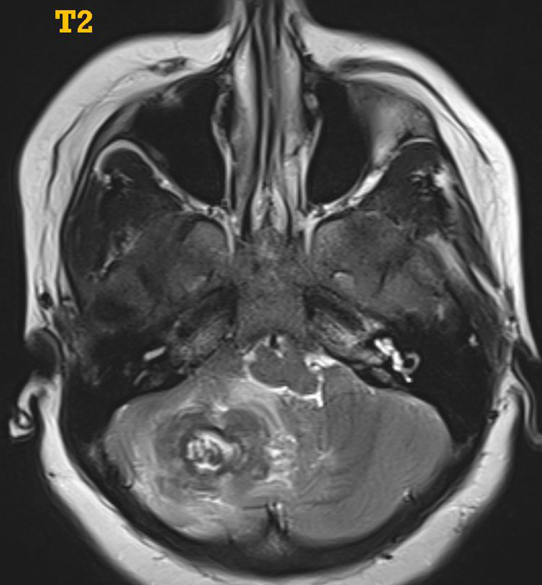

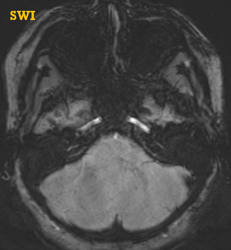
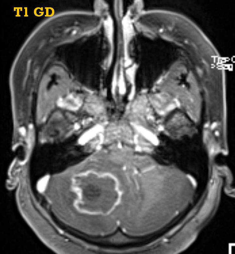
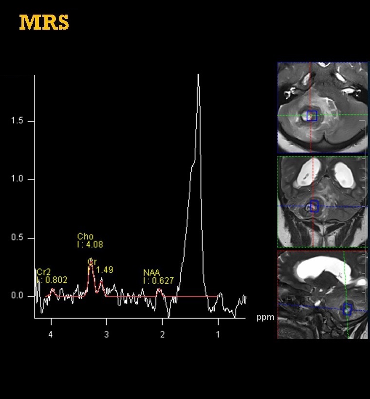
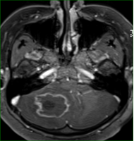
Lobulated T2 heterogeneous signal lesion in the right cerebellar hemisphere showing peripheral rim enhancement. No diffusion restriction or hemorrhage. Intermediate TE MRS demonstrates prominent lipid peak in the central part of the lesion.
Diagnosis:
Giant Intracranial Tuberculoma
Discussion:
- Solitary intracranial tuberculoma can cause diagnostic difficulties with other intracranial focal lesions like the healing stage of neurocysticercosis, fungal granulomas, chronic pyogenic brain abscesses, lymphomas, gliomas, and metastases.
- Key Diagnostic Features:
Depending on its stage of maturation, a tuberculoma’s appearance varies on MRI, i.e., whether noncaseating, caseating with a solid center, or caseating with a liquid center (Ref image below). - On MT TIW imaging, cellular components of the lesion (i.e wall) appear brighter and relatively specific for the disease. MR spectroscopy mostly demonstrates a prominent lipid peak.

Dr. Sriram Patwari
MD, PDCC (Neuroradiology)
Consultant Radiology, Co-lead Neuroradiology
Manipal Hospitals Radiology Group
Dr. Surendra K L
DMRD, DNB, FRCR, EDIR
Junior Consultant- Radiology
Manipal Hospitals Radiology Group