
A 69 year old lady with abdominal pain since 3 days.
A 69 year old lady with abdominal pain since 3 days.


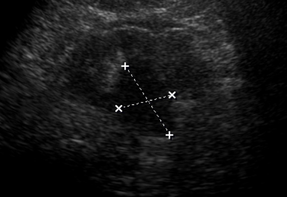
- Ultrasound image demonstrates a well defined hypoechoic lesion in the right kidney.
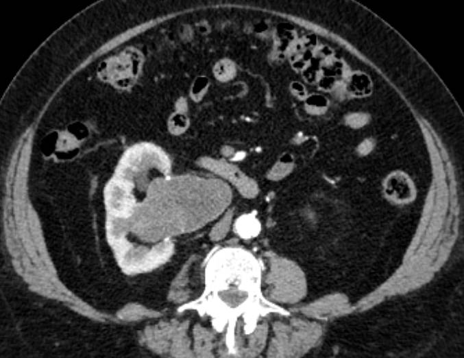
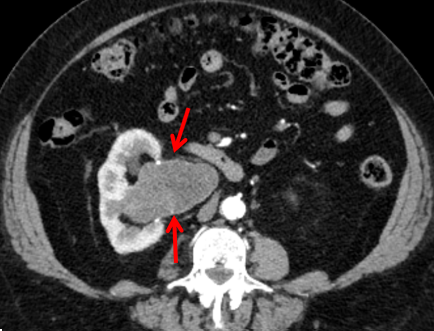
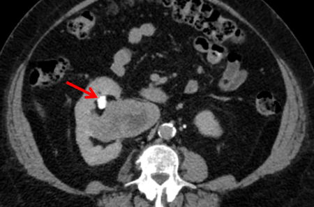
- CECT abdomen, arterial phase axial image demonstrates an exophytic, well circumscribed, lobulated, mildly enhancing soft tissue density mass in the region of the right renal pelvis.
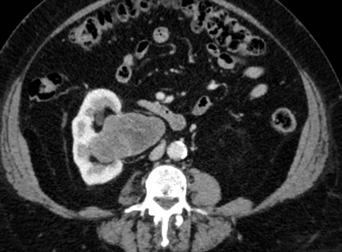
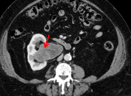
- CECT abdomen, venous and delayed phase axial images demonstrate no significant change in the degree of enhancement within the mass. The delayed phase images demonstrate that the lesion is separate from the renal pelvicalyceal system.
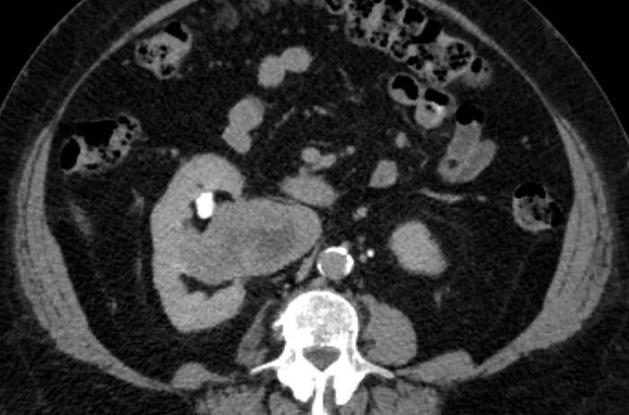
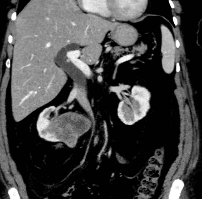
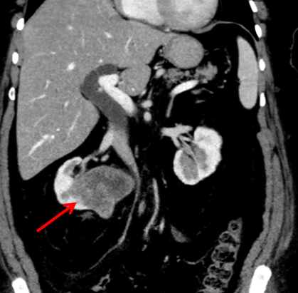
- Coronal reformatted image of CECT abdomen in the venous phase, demonstrates the parenchymal origin of the mass with extension into the renal sinus fat.
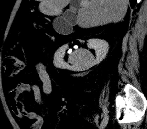
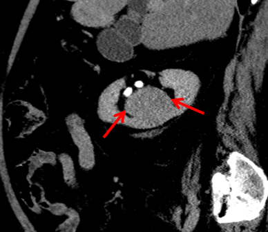
- Sagittal reformatted image of CECT abdomen in the delayed phase, demonstrates anterior displacement of the lower pole calyx by the mass with no evidence of invasion into the collecting system.
Diagnosis:
Right Renal Schwannoma
Discussion:
- Schwannomas of the kidney are rare and often incidentally detected as in our case.
- In most cases, they are seen in the renal hilum as the parasympathetic nerve fibres accompany the renal artery in the hilum. Rarely, they may arise in the parenchyma and are difficult to differentiate from renal cell carcinoma.
- On imaging, they are seen as solitary, well circumscribed masses and may demonstrate cystic degeneration and calcification, if large. Homogenous contrast enhancement is seen in the solid components.
- Imaging features are non specific and the differentials include renal cell carcinoma, transitional cell carcinoma, medullary fibroma and solitary fibrous tumour and sarcomatoid carcinoma.
- A well circumscribed renal mass in an asymptomatic patient with no evidence of vascular or pelvicalyceal sytem invasion (despite large size) should prompt the listing of a renal schwannoma in the differential diagnosis.
- Treatment is usually surgical.
References:
- A Case of Renal Schwannoma. Hee Jo Yang, Min Ho Lee, Doo Sang Kim, Hyun Ju Lee, Ji Hye Lee, Youn Soo Jeon. Korean J Urol. 2012 Dec; 53(12): 875–878.
- Mesenchymal neoplasms of the kidney in adults: Imaging spectrum with radiologic-pathologic correlation. Katabathina VS, Vikram R, Nagar AM, Tamboli P, Menias CO, Prasad SR. Radiographics. 2010;30(6):1525?1540. doi:10.1148/rg.306105517
Dr. Shilpashree G
DMRD
Radiology Resident
Manipal Hospitals Radiology Group
Dr. Anita Nagadi
MD, MRCPCH, FRCR, CCT
Senior Consultant Radiology
Manipal Hospitals Radiology Group