
30 years old male with history of low back pain and paresthesia in bilateral lower limb
30 years old male with a history of low back pain and paresthesia in bilateral lower limb
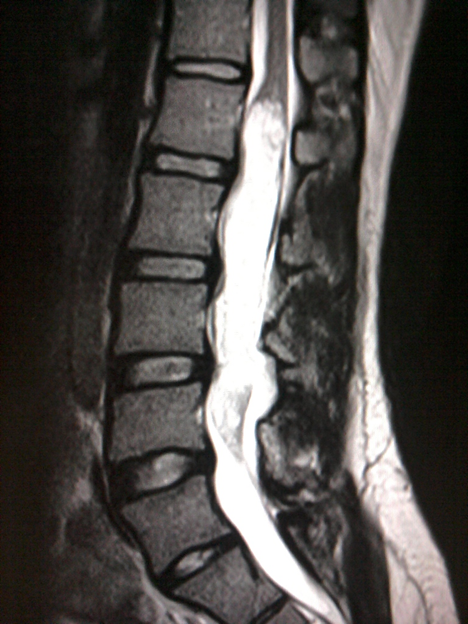
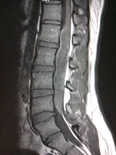
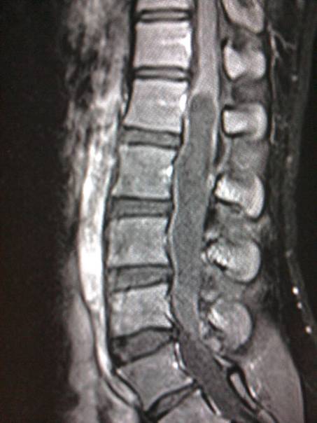
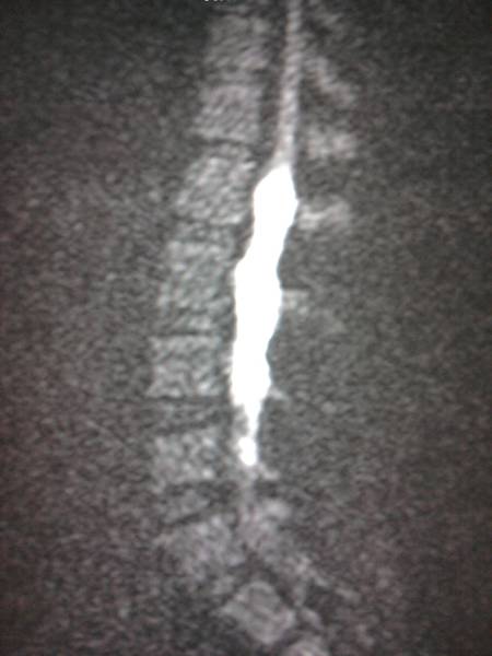
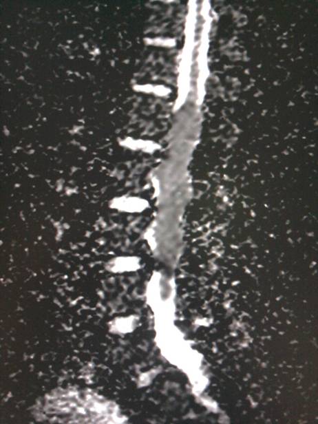
A well defined intradural T2 hyperintense (A) , T1 hypointense (B) non-enhancing (C) lesion occupying cauda equina with restriction on DWI & ADC images (D & E)
Diagnosis:
Intraspinal Epidermoid cyst
Discussion:
- Intraspinal epidermoid cysts represent less than 1% of intraspinal tumors in adults, with a higher incidence in children. They are usually extramedullary but rarely can be intramedullary, and can be congenital or acquired
- Congenital epidermoids often have associated epidermal defects, such as spina bifida and hemivertebrae, whereas acquired lesions lack osseous abnormalities. Approximately 40% of intraspinal epidermoid cysts are acquired and are considered to be a late complication of lumbar puncture.
- Clinically intraspinal epidermoid cysts may be asymptomatic and discovered incidentally. If symptomatic, motor disturbances, pain, sensory disturbances, and bowel or bladder dysfunction may be present.
- Key Diagnostic Features: Although the signal intensity of epidermoid cyst varies, it is typically iso- or slightly hyperintense compared with that of CSF on all sequences, shows no contrast enhancement, and restricts on DWI.
- DDx: Arachnoid cyst, Dermoid cyst, Cystic neoplasm
- Treatment: Surgical excision
Dr. Sriram Patwari
MD, PDCC (Neuroradiology)
Consultant Radiology, Co-lead Neuroradiology
Manipal Hospitals Radiology Group