
38 years old lady with end stage renal disease on hemodialysis, presenting with altered sensorium
38 years old lady with end-stage renal disease on hemodialysis, presenting with altered sensorium.
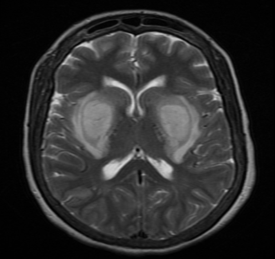
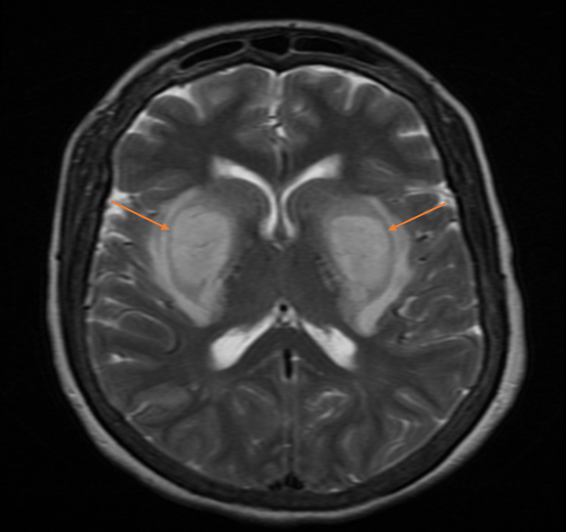
- Axial T2 and FLAIR demonstrate swollen hyperintense bilateral lentiform nucleus.
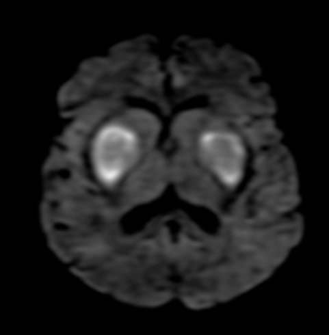
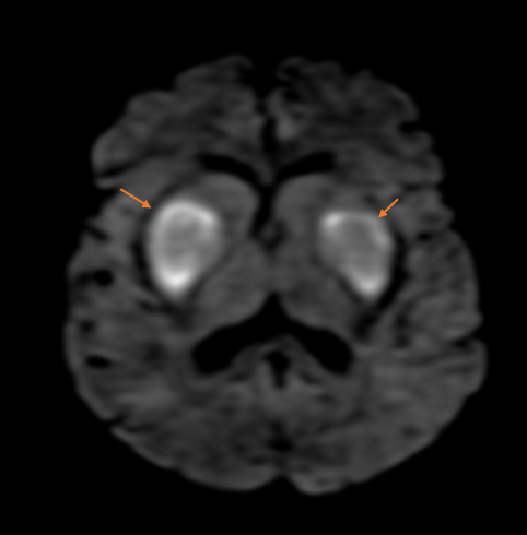
- Axial DWI demonstrates hyperintensity with the dark signal on ADC (not shown) suggesting cytotoxic edema.
- Axial GRE did not demonstrate any blood products.
DIAGNOSIS:
Uremic encephalopathy with Lentiform Fork sign
DISCUSSION:
- Uremic complications of CNS may result from either multiple metabolic derangements associated with end-stage renal disease or dialysis procedures.
- The clinical features include altered mentation, headache, nausea and vomiting, myoclonus, tremor, and focal or generalized seizures
- Key Diagnostic Features: MRI demonstrates bilateral symmetrical swollen lentiform nucleus with T2/ FLAIR hyperintensity with the margins of lentiform nucleus delineated by hyperintensity resembling fork. Mostly this represents vasogenic edema, however, in our case diffusion restriction is seen suggesting cytotoxic edema with irreversible neuronal damage. Associated diffuse cerebral edema can also be seen
- The Lentiform Fork sign is a unique MRI feature that is seen not only in patients with uremic encephalopathy but also in other conditions that result in metabolic acidosis
Dr. Sriram S Patwari MD, PDCC.
Consultant Radiologist and Co-lead Neuroradiology
Manipal Hospitals Radiology Group.