
44 year-old gentleman presented with cough and breathlessness since 2 months. Quit smoking 1 year back. Keeps cats as pets.
44 year-old gentleman presented with cough and breathlessness since 2 months. Quit smoking 1 year back. Keeps cats as pets.
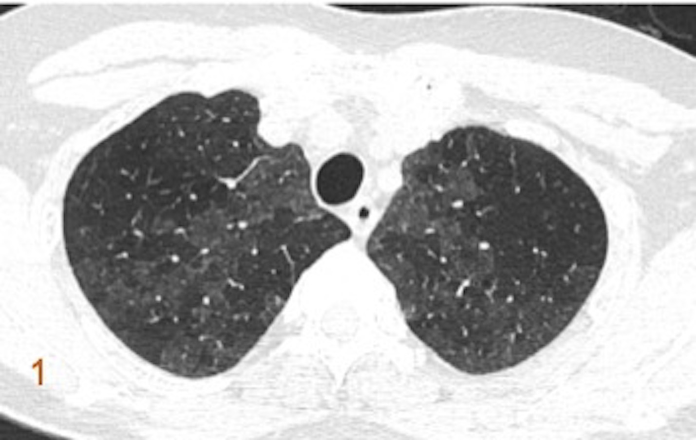
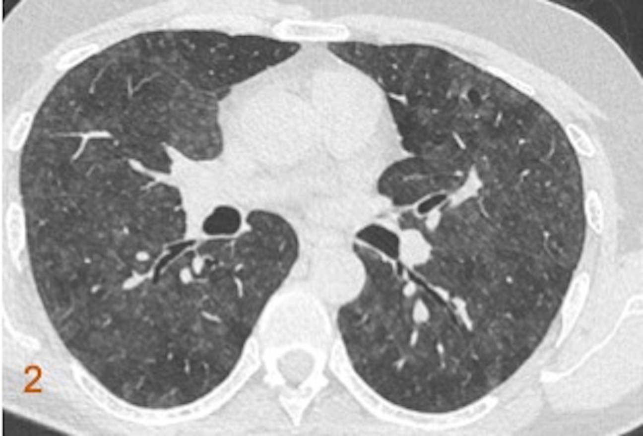


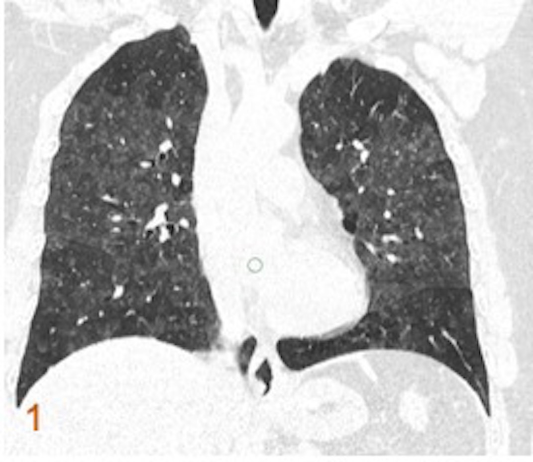
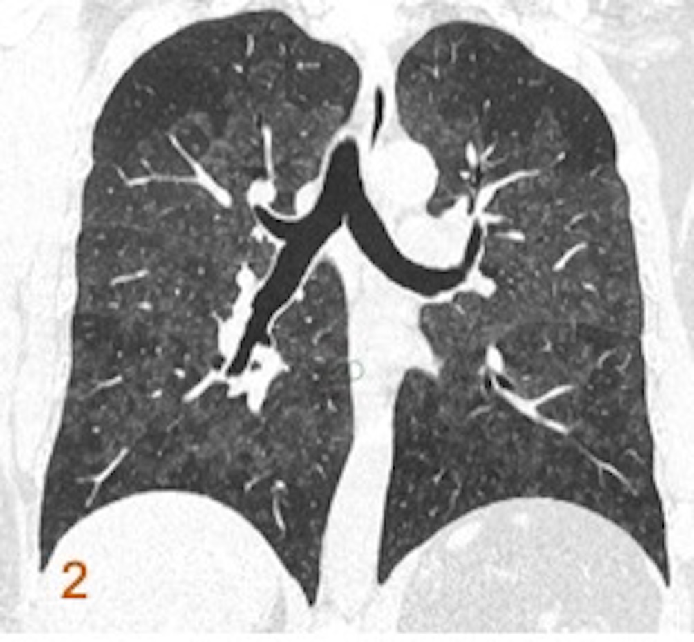
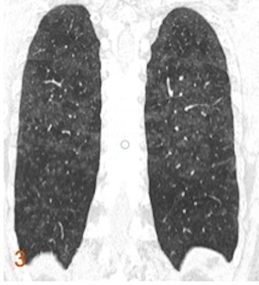
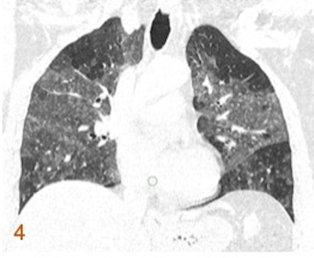

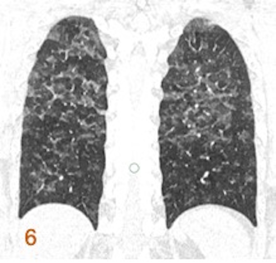
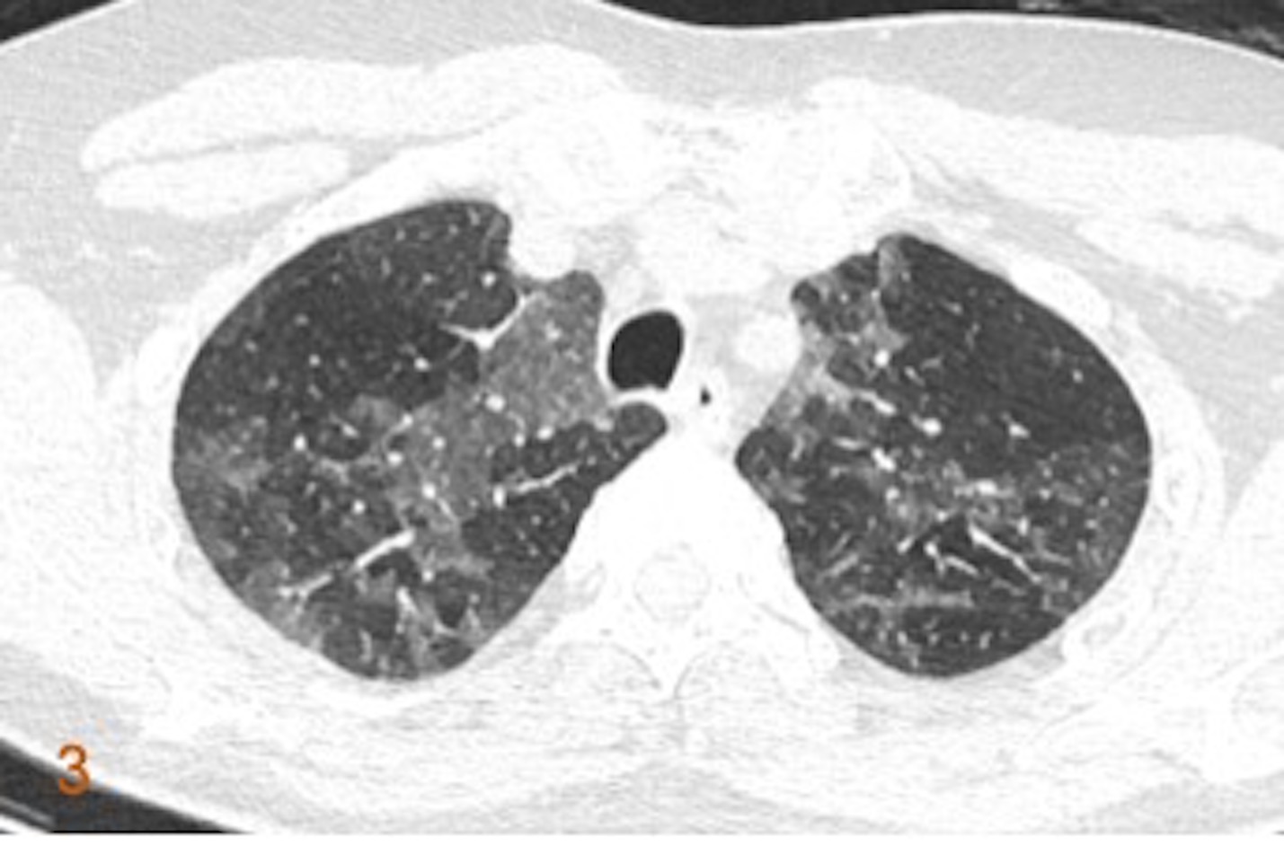
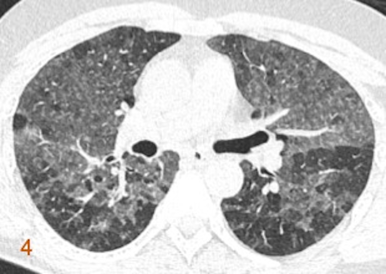
Fig 1,2 (Top row). Axial inspiratory images. Extensive, bilateral, symmetric ground-glass opacities, poorly defined centrilobular nodules, and mosaic attenuation on inspiratory images. Patchy areas of subtle traction bronchiectasis.
Fig 3,4 (Bottom row). Axial expiratory images (corresponding). Lobular areas of decreased attenuation with air trapping on expiratory CT.
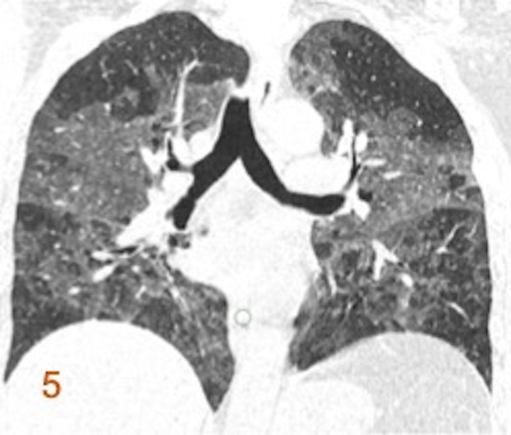
Fig 1-3 (Top row). Coronal inspiratory images. Extensive, bilateral, symmetric ground-glass opacities, poorly defined centrilobular nodules, and mosaic attenuation on inspiratory images. Patchy areas of subtle traction bronchiectasis.
Fig 4-6 (Bottom row). Coronal expiratory images (corresponding). Lobular areas of decreased attenuation with air trapping on expiratory CT.


Diagnosis:
Hypersensitivity pneumonitis, Cluster 1.
Discussion:
- Hypersensitivity pneumonitis is a chronic granulomatous interstitial lung disease caused by inhalation/ exposure to antigens such as hay, bird proteins and molds.
- Imaging Findings on HRCT Chest:
- In Acute/ inflammatory HP: Upper and mid zone predominance. Ground glass and centrilobular ground glass nodules, areas of mosaic attenuation with lobular areas of low attenuation and expiratory air trapping. Generally spared lung bases. Head cheese sign is characterized by presence of lobular well demarcated areas of normal lung, low and high attenuation lung.
- In Fibrotic HP: Can have lower lobe and subpleural distribution of ground glass with reticulations, traction bronchiectasis and honeycombing.
- Differential diagnosis:
Fibrotic NSIP and IPF rarely have centrilobular nodules and lobular areas of low attenuation.
References:
- Silva CI, Churg A, Mu?ller NL. Hypersensitivity pneumonitis: spectrum of high-resolution CT and pathologic findings. American Journal of Roentgenology. 2007 Feb;188(2):334-44.
- Selman M, Pardo A, King Jr TE. Hypersensitivity pneumonitis: insights in diagnosis and pathobiology. American journal of respiratory and critical care medicine. 2012 Aug 15;186(4):314-24.
Dr. Deepali Saxena
DNB, Fellowship Cardiothoracic Imaging (USA)
Lead Cardiac Imaging
Columbia Asia Radiology Group