
65 year-old chronic smoker presented with breathlessness
65 year-old chronic smoker presented with breathlessness.
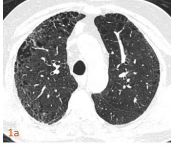

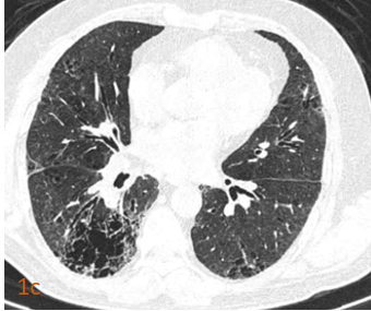
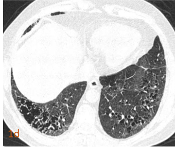
- Fig 1 (a,b,c,d). Axial HRCT Chest inspiratory images. Bizarre shaped, thin walled, irregular sized clustered cysts, sparing the subpleural parenchyma. Few reticulations noted.
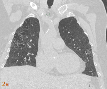
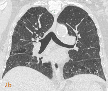
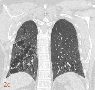
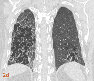
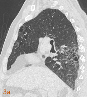
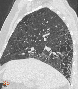
- Fig 2 (a,b,c,d). Coronal HRCT Chest inspiratory images and Fig 3 (a,b) Sagittal HRCT Chest inspiratory images. Bizarre shaped, thin walled, irregular sized clustered cysts, sparing the subpleural parenchyma. Few reticulations noted.
Diagnosis:
Smoking Related Interstitial Fibrosis (SRIF) with combined emphysema.
Discussion:
- Hallmarks of smoking related interstitial fibrosis (SRIF) with combned emphysema, on HRCT chest are:
- Thin walled clustered cysts that are bizarre in shape and irregular in size
- Ground glass opacities
- Reticulations.
- Subpleural parenchyma is less involved.
- As the name suggests, etiology is smoking.
- It represents a spectrum of changes that occur in smoking related interstitial lung diseases.
- To D/D from thin walled honeycombing in UIP, SRIF with emphysema has:
- Thin walled clustered cysts that are irregular in size and bizarre in shape c.f., rounded, uniform layered cysts.
- Subpleural parenchyma is spared c.f., involves subpleural parenchyma.
References:
- Otani H, Tanaka T, Murata K, Fukuoka J, Nitta N, Nagatani Y, Sonoda A, Takahashi M. Smoking-related interstitial fibrosis combined with pulmonary emphysema: computed tomography-pathologic correlative study using lobectomy specimens. International journal of chronic obstructive pulmonary disease. 2016;11:1521.
Dr. Deepali Saxena,
DNB, Fellowship Cardiothoracic Imaging (USA)
Lead Cardiac Imaging
Manipal Hospitals Radiology Group.