
The 73-year-old female is experiencing pain on the lateral aspect of her ankle
- 73-year-old female
- Pain on the lateral aspect of the ankle
- No history of trauma

FINDINGS
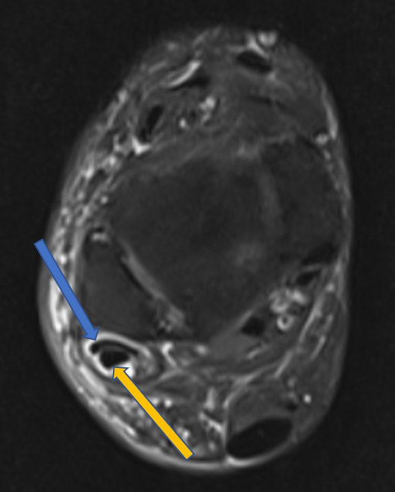
- Coronal and axial PDF images distal to the level of lateral malleolus demonstrate soft tissue hyperintensity surrounding the peroneal tendons (Blue arrows).
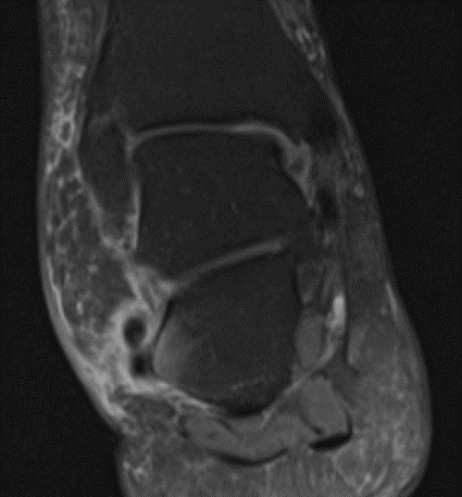
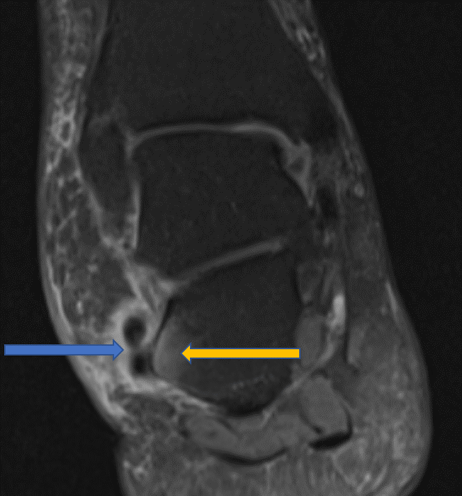
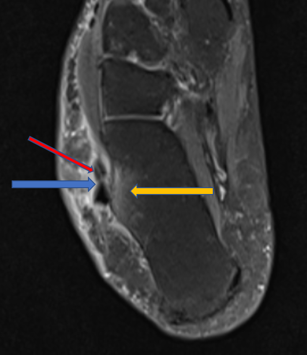
- Prominent peroneal tubercle of the calcaneus with bone marrow edema (Yellow arrows) adjacent to the peroneal tendons. Linear hyperintensity is seen within the peroneus brevis tendon (red arrow) in Fig 2.
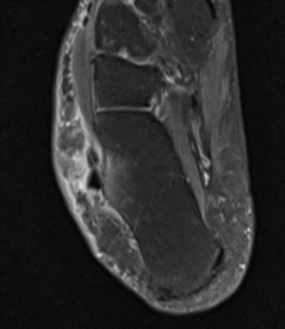
- Fluid and soft tissue edema surrounding peroneus longus (yellow arrow) and peroneus brevis (blue arrow) tendons behind and distal to the level of the lateral malleolus.
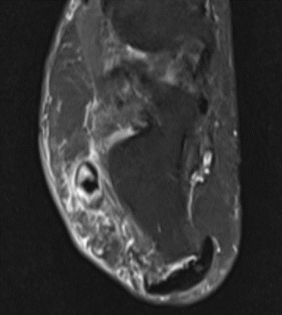
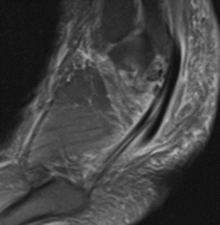
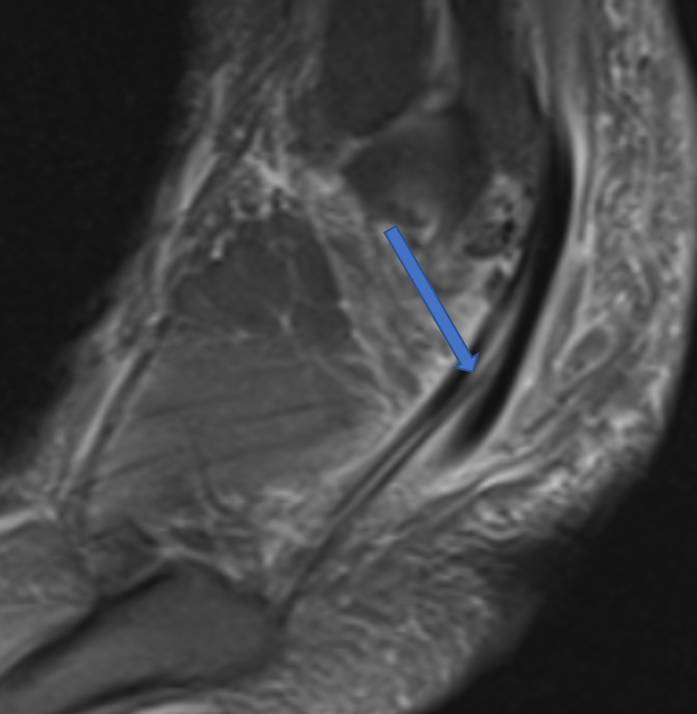
- Intrasubstance high signal with inverted “U” shaped configuration of peroneus brevis tendon partially encircling the peroneus longus tendon in figs 3 and 4. Linear intra-substance hyperintensity through the length of the peroneus brevis in Fig 5.

Diagnosis:
- Tenosynovitis of peroneal tendons with a longitudinal split tear of the peroneus brevis tendon.
- Prominent peroneal tubercle of calcaneus with bone marrow edema.
Features are suggestive of peroneal tubercle friction syndrome
DISCUSSION:
Relevant anatomy:
- -Peroneal tendons are paired tendons in posterolateral aspect of ankle and foot
- -Both tendons are enclosed within a common tendon sheath proximal to the level of tip of lateral malleolus.
- -Superior peroneal retinaculum and lateral malleolus forms a fibro-osseous tunnel that stabilizes peroneal tendons in retro malleolar region.
- -More distally, the tendons are enclosed within separate tendon sheaths separated by the peroneal tubercle of calcaneus. Inferior peroneal retinaculum forms two separate fibro-osseous tunnels fore each tendon at the level of peroneal tubercle.
- -At the level of ankle, peroneus brevis tendon lies closer to malleolus than longus.
- -At the level of peroneal tubercle, the brevis tendon is anterosuperior and the longus tendon is posteroinferior.
Peroneal tubercle friction syndrome:
- -Prominent peroneal tubercle is most often congenital and also can be post-traumatic.
- -Recurrent friction/chronic irritation of peroneal tendons and tendon sheath as they glide over hypertrophic peroneal tubercle.
- -Spectrum of abnormality can range from tendinosis, tenosynovitis, partial tendon tear, or complete tendon tear.
- -Bone marrow edema in peroneal tubercle and adjacent calcaneus
Peroneus brevis split tear: MR appearances
- -Longitudinal intra-substance high signal within the tendon.
- -Abnormal inverted “U” shaped tendon or “Boomerang” shaped brevis tendon enclosing longus tendon.
- -Separation of brevis tendon into two smaller and separate hemi tendons with longus tendons interposed between them.
- -Focal thinning of tendon/contour abnormality of tendon confirmed in two separate imaging planes.
- -Complete discontinuity of tendon.
The differential diagnosis for peroneus brevis split tears:
(a)Bifurcated peroneus brevis tendon: differentiated by identifying two separate myotendinous junctions proximally.
(b)Peroneus quadratus (accessory muscle) inserted into brevis tendon.
Dr. Dayanand Sagar
G, MD
Consultant Radiologist, MGRG
