
The 37-year-old male has been experiencing swelling in his right foot for the past five years
- 37 year, male
- Swelling of the right foot for 5 years
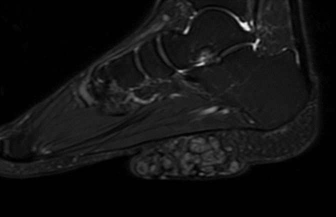
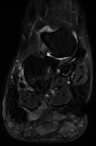
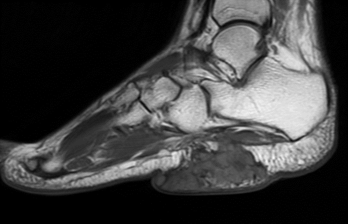
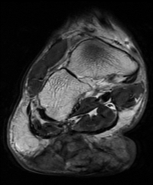
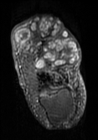
- Irregular T1 iso-hypo intense and T2 heterogeneously hyperintense mass in the subcutaneous plane of the mid-posterior foot with lobulated margins (blue arrow).
- The mass demonstrates nodular T2 hyper-intense areas with a central dot (Dot in circle sign) interspersed with T2 hypo intense fibrous areas (Orange arrow)
- Mass insinuating between flexor digitorum brevis and abductor digit minimi without obvious muscles invasion (yellow arrow)
- No evidence of calcifications within the mass. No bony lesions (purple arrow)
Diagnosis: Madura foot
DISCUSSION:
- Mycetoma is a chronic granulomatous fungal infection, endemic in the tropics, mainly Africa, Mexico and India.
- It is named after Madurai in India, where it was originally described in 1842.
- It commonly affects the feet, hands, back, and gluteal region.
- It is caused by either actinomycetes or eumycetes group of fungi.
Clinical perspective:
- It typically presents in farmers who walk barefoot in dry, dusty conditions.
- Minor trauma allows pathogens to enter the skin from the soil and gradually form discharging granulomas with subsequent involvement and destruction of underlying bones.
- It is important to differentiate between actinomycetoma and eumycetoma because of the different responses to treatment.
- Meticulous diagnosis of the fungus can save the weight-bearing function of the foot as well as circumvent the need for surgical amputation
Treatment/Outcome:
- Surgical debridement followed by prolonged antibiotic therapy for several months is required for actinomycetoma.
- Eumycetomas are only partially responsive to anti-fungal therapy but can be treated by surgery due to their normally well circumscribed nature.
- Surgery in combination with azole treatment is the recommended regime for small eumycetoma lesions in the extremities. Amputation may be required in recurrent cases
Imaging perspective :
- Radiographs/Computed tomography: X-ray/CT shows changes of chronic osteomyelitis with soft tissue involvement, sclerosis, cavitation, cortical erosion and destruction of underlying bones.
- Conventional radiographs/Computed tomography are used to determine whether bones are affected and to identify the limits of lesions.
- MRI: MRI is useful for visualizing soft tissue involvement and bone destruction.
- Multiple small spherical hyperintense granulomatous lesions separated by tissues of low signal intensity appear, with hypointense foci of fungal hyphae, consistent with “Dot-in-circle”, which makes this appearance characteristic of Mycetoma. This feature is also noted on ultrasound.
- Actinomycetoma more often delineate soft tissue microabscesses, bony periosteal reaction and reactive sclerosis, while eumycetoma frequently exhibit soft tissue macroabscesses with bone cavitation.
- However, culture studies remain the gold standard for species identification.


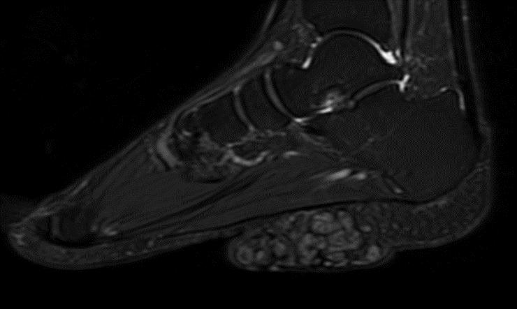
Axial and Sagittal T2-weighted MR image of the foot showing conglomerates of discrete small round hyperintense lesions with peripheral hypointense rim and central hypo intensities within (arrows) in calcaneum and the dorsal and plantar aspects of medial aspect of distal right foot, few lesions are noted to infiltrate into the muscles.
Teaching points:
- Typical clinical history with clear radiological signs can lead to early diagnosis of Madura foot and prevent deformity/remodeling.
- It is important to identify the causative species for implementing the correct line of treatment.
- The dot-in-circle sign is characteristic of maduramycosis on MRI and ultrasound.
REFERENCE:
- Cherian RS, et al. (2009) The “dot-in-circle” sign — a characteristic MRI finding in mycetoma foot: a report of three cases. Br J Radiol Aug;82(980):662-5 (PMID: 19221181)
- Sen A, et al. (2011) Case report: Dot-in-circle sign – An MRI and USG sign for. Indian J Radiol Imaging Oct;21(4):264-6 (PMID: 22223936)
- Kumar J, et al. (2007) The dot-in-circle sign of mycetoma on MRI. Diagn Interv Radiol Dec;13(4):193-5 (PMID: 18092291)
- Rao AG. (2016) Mycetoma. Indian Dermatol Online J Jul-Aug;7(4):353-5 (PMID: 27559532)
Dr. Srinivas P
Cross-sectional Fellow
Manipal Hospital Radiology Group (MHRG)
Manipal Hospital, Bengaluru
Dr. Dayanand Sagar
Consultant Radiologist.
Manipal Hospital Radiology Group (MHRG)
Manipal Hospital, Bengaluru.
