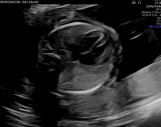
A three week old male infant presents with respiratory distress and sepsis since birth with suspected CPAM on antenatal ultrasound
A three week old male infant presents with respiratory distress and sepsis since birth with suspected CPAM on antenatal ultrasound
FINDINGS:
- A: Antenatal USG showing echogenic lesion in right lung.
- B: Patchy opacity in right middle zone.
- C: MinIP image showing Hyperlucent right upper lobe anterior and apical segment (arrow) with multiple scattered cystic areas within (arrow head).
- D: Narrowing of segmental and subsegmental bronchioles with paucity of vessels (arrow).
- E: Compensatory hypertrophy of posterior segment of right upper lobe (arrow).
DIAGNOSIS:
- Type III Congenital pulmonary airway malformation.
DISCUSSION:
Congenital pulmonary airway malformations (CPAM) are multicystic masses of segmental lung tissue with abnormal bronchial proliferation. CPAMs are considered part of the spectrum of bronchopulmonary foregut malformations.
Associations
- hybrid lesion: i.e. CPAM and pulmonary sequestration.
- renal agenesis.
- Polyhydramnios
- hydrops fetalis
- congenital cardiac anomalies
- lung malignancy
Clinical presentation:
- The diagnosis is usually either made on antenatal ultrasound, or in the neonatal period on the investigation of progressive respiratory distress. If large, they may cause pulmonary hypoplasia, with a resultant poor prognosis.
- In cases where the abnormality is small, the diagnosis may not be made for many years or even until adulthood. When it does become apparent, it is usually as a result of recurrent chest infection.

Radiographic features:
- The appearance of CPAM will vary depending on the type.
Plain radiograph:
- Chest radiographs in type I and II CPAM may demonstrate a multicystic lesion. Large lesions may cause a mass effect with resultant mediastinal shift, depression, and even inversion of the diaphragm. In the early neonatal period, the cysts may be completely or partially fluid-filled, in which case the lesion may appear solid or with air-fluid levels.
Ultrasound:
- CPAM appears as an isolated cystic or solid intrathoracic mass. A solid thoracic mass is usually indicative of a type III CPAM and is typically hyperechoic. There can be a mass effect where the heart may appear displaced to the opposite side.
- Hydrops fetalis and polyhydramnios may develop and may be detected on antenatal ultrasound as ancillary sonographic features.
CT:
- CT has a number of roles in the management of CPAM. First, it more accurately delineates the location and extent of the lesion. CT angiography is able to identify systemic arterial supply if present.
- Appearance reflects the underlying type, with features of hyperlucency, variable sized cyst depending on type and associated complication. A type III lesion can appear as a consolidation.
Complications:
Potential postnatal complications include:
- Recurrent pneumothorax
- Hemopneumothorax
- Pyopneumothorax
- Malignancies
Differentials:
Bronchogenic cyst
- unilocular
- does not usually communicate with the bronchial tree, and are therefore typically not air-filled
Pulmonary sequestration
- systemic arterial supply
- hybrid lesions may present with both CPAM and sequestration features (see above)
Congenital diaphragmatic herniation
Congenital lobar emphysema (congenital lobar overinflation)
- hyperlucent and hyperinflated lung segment
- no cystic or solid components
References:
- Biyyam, D.R. et al. (2010) ‘Congenital lung abnormalities: Embryologic features, prenatal diagnosis, and postnatal radiologic-pathologic correlation’, RadioGraphics, 30(6), pp. 1721–1738. doi:10.1148/rg.306105508.
- Congenital pulmonary airway malformation | radiology reference article | radiopaedia.org. Available at: https://radiopaedia.org/articles/congenital-pulmonary-airway-malformation (Accessed: 24 November 2024).
Dr DEEPT H V
Senior Consultant Radiologist
Manipal hospitals, Yeshwantpur
DR VASANTH KUMAR
Cross Section Fellow
Manipal Hospitals, Yeshwantpur


