

A 9 year old female presented with abdominal discomfort.
A 9 year old female presented with abdominal discomfort.
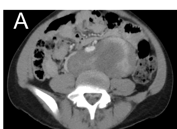
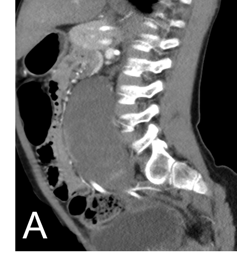
- A. FINDINGS – CECT ABDOMEN AND PELVIS
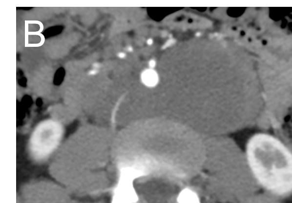
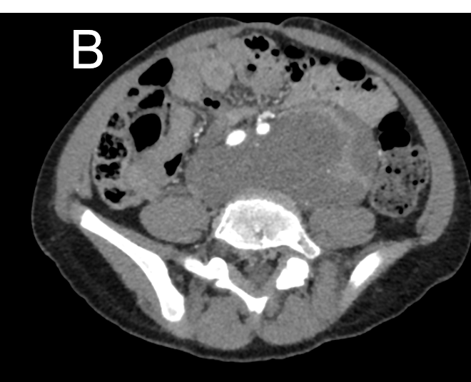
- B. FINDINGS – CECT ABDOMEN AND PELVIS
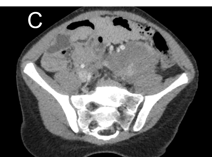
- C. FINDINGS – CECT ABDOMEN AND PELVIS
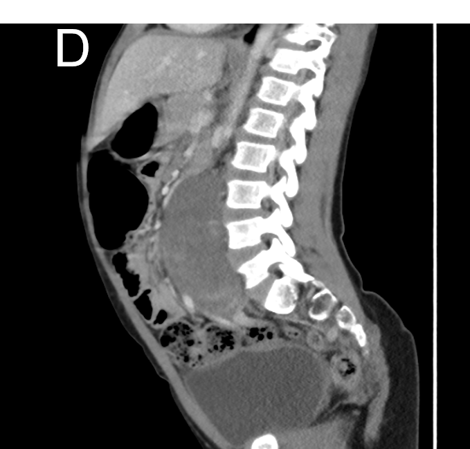
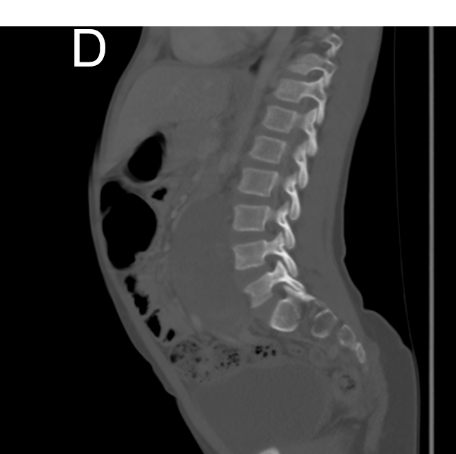
- D. FINDINGS – CECT ABDOMEN AND PELVIS
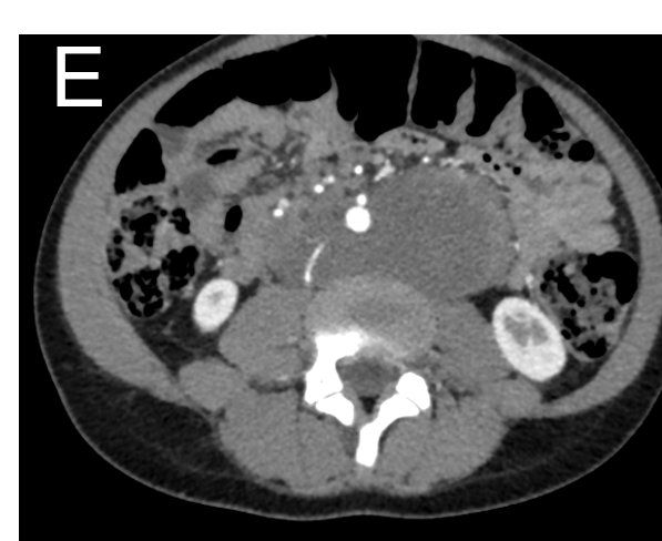
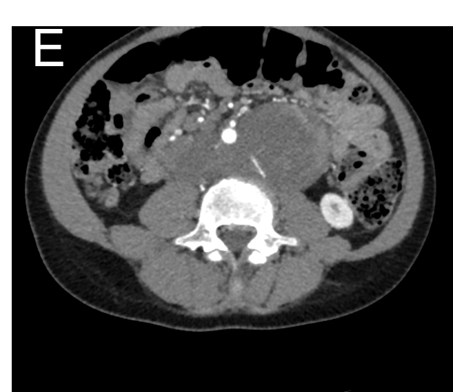
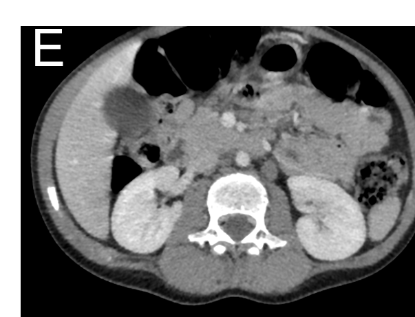
- E. FINDINGS – CECT ABDOMEN AND PELVIS
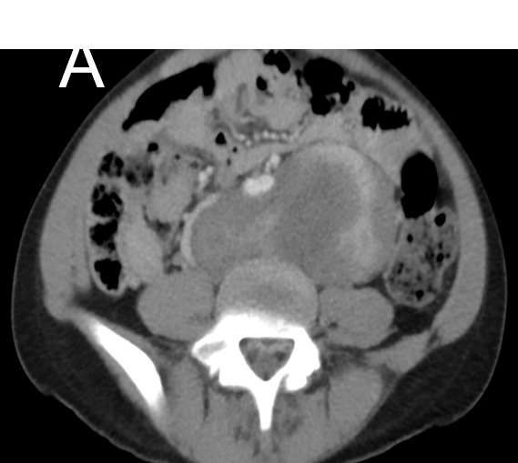
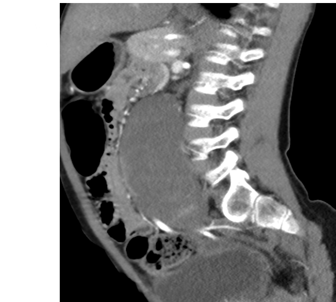
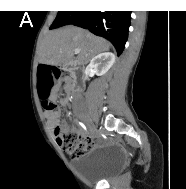
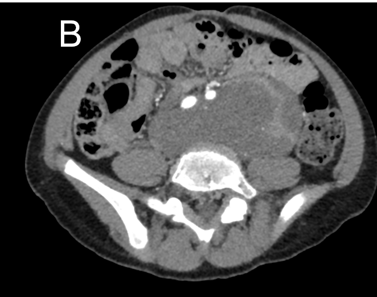
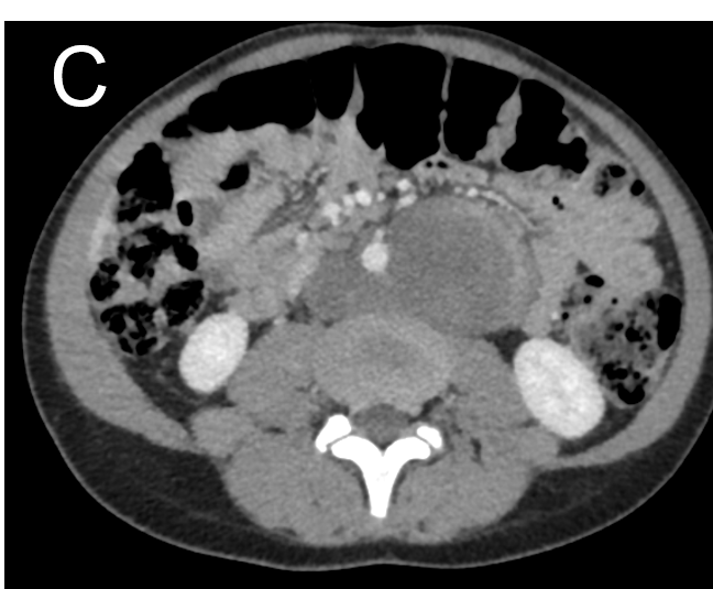
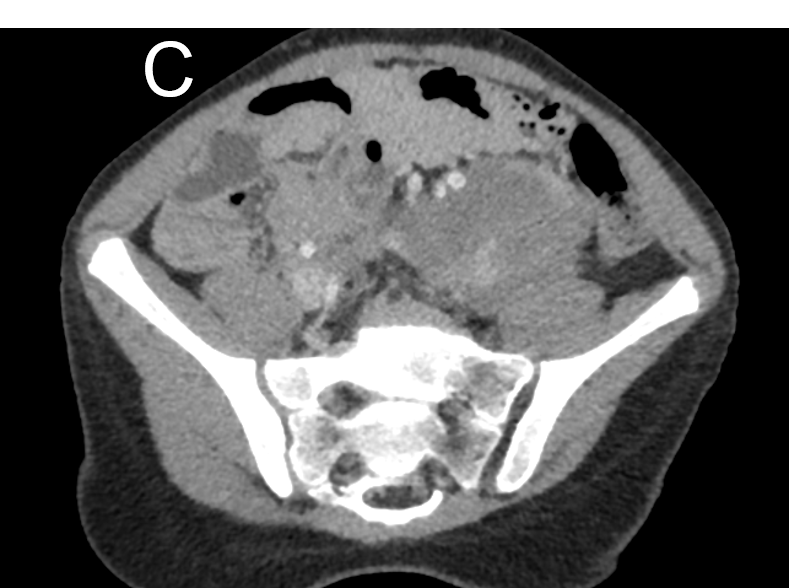

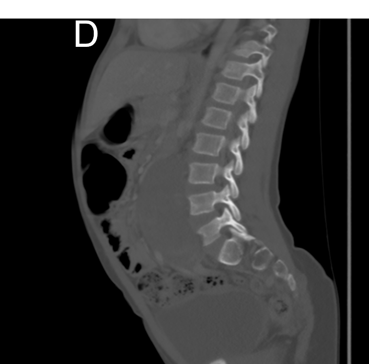
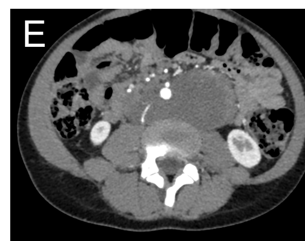
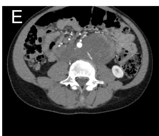
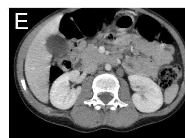
Large well defined lobulated heterogeneously enhancing soft tissue lesion in the retroperitoneum , situated posterior to the aorta. Extends superiorly up to L2-L3 intervertebral disc space & inferiorly up to L5-S1 intervertebral disc space.
- A. Antero-inferiorly, - Encasing (<270 degree) the infra renal aorta, displacing it anteriorly without infiltration or thrombosis. - Abutment (<180 degree) and displacement of bilateral common iliac arteries and left proximal external iliac artery. - Displacing the small and large bowel loops anteriorly without infiltration.
- B. Antero-inferiorly, - Encasing (<270 degree) the infra renal aorta, displacing it anteriorly without infiltration or thrombosis. - Abutment (<180 degree) and displacement of bilateral common iliac arteries and left proximal external iliac artery. - Displacing the small and large bowel loops anteriorly without infiltration.
- C. Right laterally, -Abutting (<180 degree) the infra renal IVC, causing significant compression and displacement without infiltration or thrombosis. -Near complete encasement with significant compression of distal left proximal common iliac vein.
- D. Posteriorly abutting the anterior surface of L4 and L5 vertebral bodies and adjoining intervertebral disc space, without infiltration or bony erosion or extension into neural foramina.
- E. Third & fourth lumbar arteries course through the lesion bilaterally. Few prominent nodes in left para aortic and aorto caval regions.

Radiological diagnosis : Ganglioneuroma Patient underwent CT guided biopsy followed by surgical excision HPE- Ganglioneuroma (Ki67% - 1% and mature ganglion cells)
- Fully differentiated neuronal tumors that do not contain immature elements and potentially occur anywhere along with the peripheral autonomic ganglion sites
- Children (mean age 7 years),slight female predominance
- Usually asymptomatic
CT
- Well-defined hypodense masses, homogenous.
- Punctate calcifications (33%)
- Slight or moderate progressive enhancement
MR
- T1- hypointense, T2- heterogeneous hyperintense, diffusion restriction
Whorled sign- microscopic interlacing patterns of Schwann cells and collagen fibers within the tumor
Vessel encasement sign- extend along the inter-vessel space and partially or wholly surround major blood vessel
DD
- Neuroblastoma
- Ganglioneuroblastoma
- Schwannoma
- Lymphangioma
Neuroblastoma- more aggressive and occur in younger patients (2 years)
NB and GNB
- Irregularly, unencapsulated lesions and tend to be inhomogeneous
- Invade adjacent organs and adjacent vessels and tend to metastasize to bone, bone marrow, liver, and lymph nodes
- Regional invasion of paraspinal musculature and psoas may occur, and invasion of the neural foramen into the epidural space
Schwannomas
- Majority of neurogenic retroperitoneum neoplasms
- 2nd to 5th decades of life
- Encapsulated masses and extend along the nerve course
- Large schwannomas may present prominent cystic degeneration and calcification
- Target-like appearance - peripheral high-signal intensity and central low-signal intensity, on T2
Retroperitoneal lymphangiomas
- Any age and are common in males
- Unilocular or multilocular thin-walled cysts with no enhancement
Management- surgery
REFERENCES
- Pacella G, Brunese MC, Donnarumma F, Barrassi M, Bellifemine F, Sciaudone G, Vallone G, Guerra G, Sallustio G. Imaging of Ganglioneuroma: A Literature Review and a Case of in an Adolescent Girl. Diagnostics (Basel). 2023 Jun 27;13(13):2190. doi: 10.3390/diagnostics13132190. PMID: 37443583; PMCID: PMC10341194.
- Xiao J, Zhao Z, Li B, Zhang T. Primary Retroperitoneal Ganglioneuroma: A Retrospective Cohort Study. Front Surg. 2021 May 21;8:642451. doi: 10.3389/fsurg.2021.642451. PMID: 34095202; PMCID: PMC8176303.
Dr MADHU KUMAR
Senior Consultant Radiologist
Manipal Hospital, Yeshwanthpur, Bengaluru.
Dr SHIKHA JOSHI
Radiology resident
Manipal Hospital, Yeshwanthpur, Bengaluru.

