A 75 year old lady with history of left breast lump for 8 years, increasing in size for 2-3 years
- A 75-year-old lady with a history of a left breast lump for 8 years, increasing in size for 2-3 years.
- The patient now presented with intermittent high-grade fever for 1 day and bloody discharge from the lower outer quadrant of the left breast.
FINDINGS
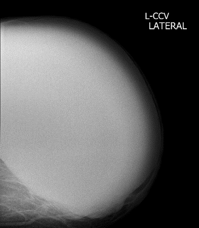
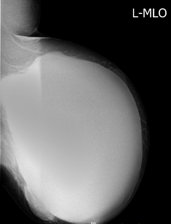
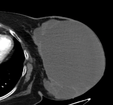
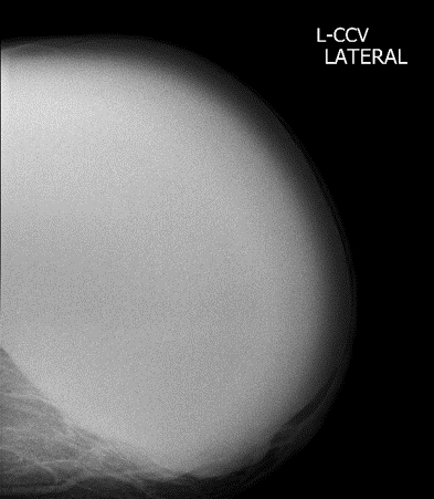
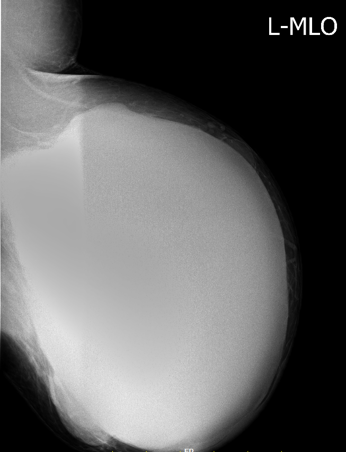
- The entire left breast is replaced by well- a defined high-density mass lesion.
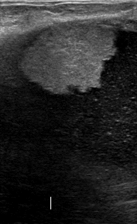
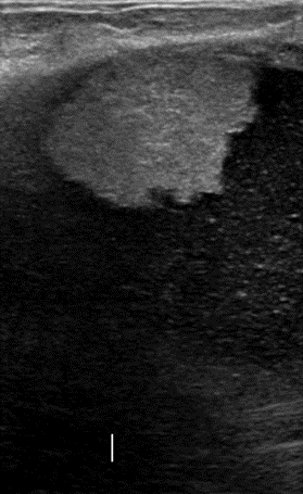
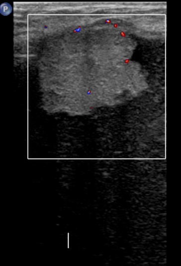
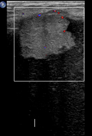
- Large complex solid cystic mass lesion with low-level echoes and associated peripheral vascularity.
- BI-RADS Assessment Category 4: Suspicious abnormality.
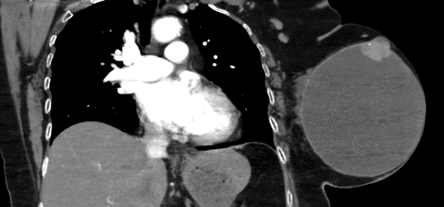
- Large well-defined cystic lesion with lobulations and few septations within in the left breast with two enhancing focal mural nodules along the superior aspect of the cystic lesion with specks of calcification within.
DIAGNOSIS:
Intra-cystic papillary carcinoma of the breast.
DISCUSSION:
- ICPC is a rare malignant tumor that accounts for 1%–2% of all breast carcinomas.
- Tumors of this type usually arise in dilated ducts, not in cysts, and are called intracystic only when the cystic component is relatively large.
- Usually does not demonstrate invasive growth.
- Usually appears in postmenopausal women, with an average age of onset of about 69.5 years.
- Tumor may be uni or multifocal and can be found as a pure form or may be associated with ductal carcinoma in situ or invasive carcinoma.
Imaging findings:
- Mammography – well-circumscribed mass with occasional satellite nodules or microcalcifications.
- Sonography – solid or complex cystic masses.
- Color Doppler sonography – intratumoral blood flow or large feeding vessels.
- Contrast-enhanced MRI – marked enhancement of cyst walls, septations and mural nodules.
Treatment:
- Mastectomy or segmental resection. Radiation therapy may also be administered.
- Sentinel lymph node biopsies or axillary dissections in the presence of nodal spread.
Prognosis:
- Pure ICPC has a slow growth rate and an excellent prognosis, with a 10-year survival rate approaching 100%.
REFERENCES
- Hernandez Rodriguez MC, LOPEZ SECADES A, Angulo JM. Best Cases from the AFIP: Intracystic Papillary Carcinoma of the Breast. RadioGraphics. 2010;30(7):2021-7.
- Wagner AE, Middleton LP, Whitman GJ. Intracystic papillary carcinoma of the breast with invasion. AJR Am J Roentgenol. 2004 Nov 1;183(5):1516.
Dr. Rashmi Jayakar Poojary
Radiology resident
Manipal Hospital, Yeshwanthpur, Bengaluru.
Dr. Anita Nagadi
Senior Consultant Radiologist
Manipal Hospital, Yeshwanthpur, Bengaluru

