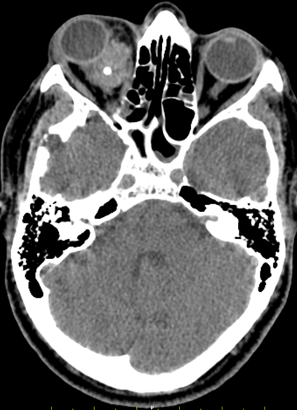
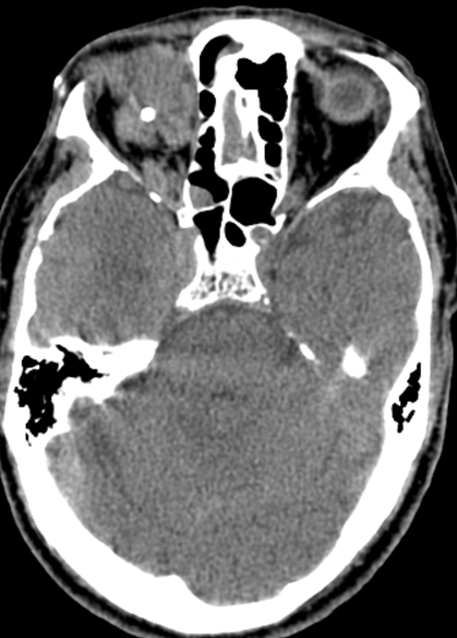
A 51 year old gentleman, with history of right eye proptosis since 15 days
A 51 year old gentleman, with history of right eye proptosis since 15 days.
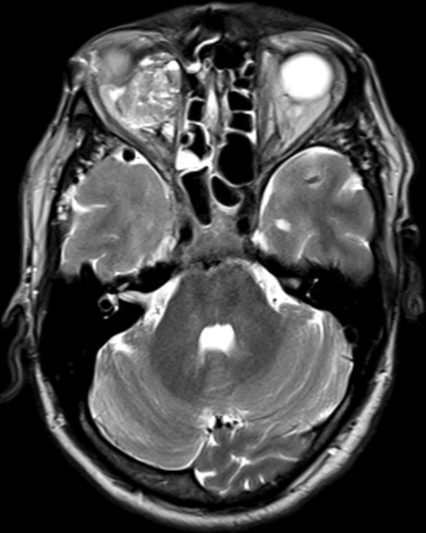
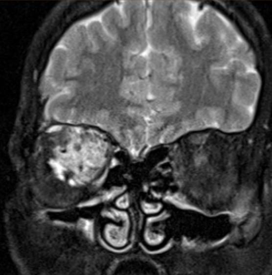
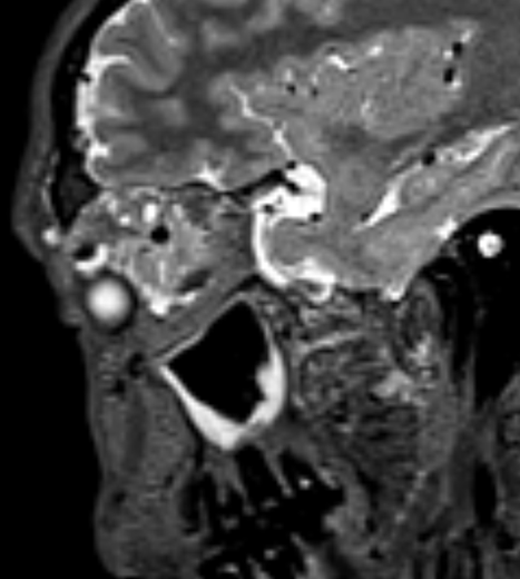
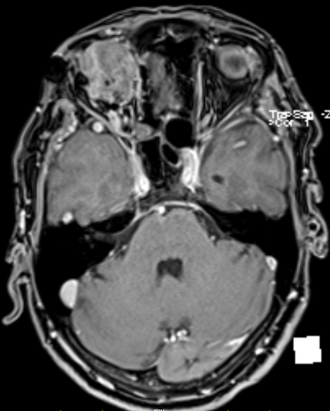
- Axial T2 weighted image demonstrates a fairly-defined, multiloculated heterogeneously T2 hyperintense lesion involving the intraconal and extraconal compartments of the right orbit. Axial T1 weighted images demonstrate multiple small cystic spaces within the lesion with heterogeneously hyper-intense contents suggestive of hemorrhagic, lymphatic, or proteinaceous content.
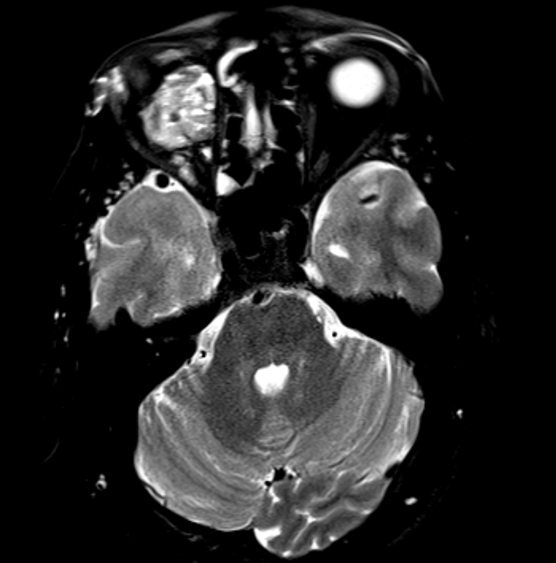
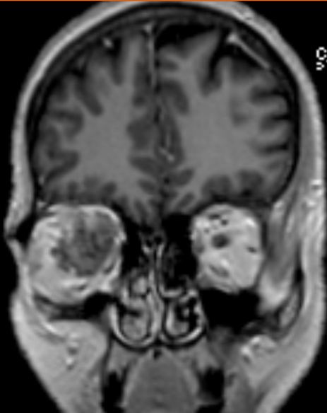
- Axial T2 fat-suppressed images demonstrate fluid-fluid levels with hypointense-dependent contents within the lesion. Coronal T2 fat-suppressed images demonstrate a hypointense pseudo capsule.
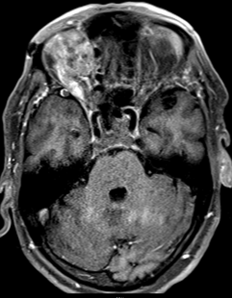
- Post-contrast T1 axial and coronal sequences demonstrate mild heterogenous enhancement of the lesion.
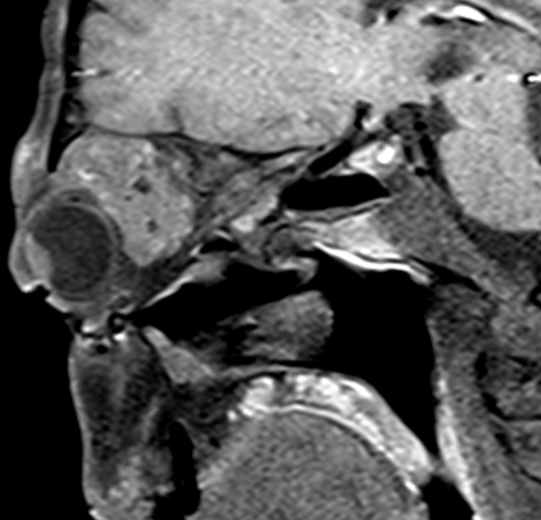
- Sagittal T2 and T1 sequences demonstrate multiple hypointense foci within the lesion. These correspond to hyperdense calcific foci on CT and are suggestive of phleboliths.
Diagnosis:
Orbital slow flow venous malformation.
Discussion:
- Also known as orbital cavernous venous malformations, these are the most common?vascular lesions of the orbits in adults.
- ?Usually presentwith a slowly growing orbital mass resulting in proptosis
Imaging:
- Most commonly located within the intra conal compartment, on the lateral aspect?.
- Usually round or oval.
- USG: Smoothly circumscribed retrobulbar lesion with regular moderate to high internal echogenicity and no demonstrable internal vascularity.
- CT: Well-circumscribed, rounded or oval soft tissue density mass, which gradually and incompletely fills in following the administration of contrast. Phleboliths can be seen.
- MRI:
T1: Isointense compared to muscle. If areas of thrombosis are present, then hyperintense regions may be visible.
T2: Hyperintense compared to muscle. Can have fluid-fluid levels with T2 hypointense pseudo capsule and T2 hypo intense phleboliths.
T1 C+ (Gd):??Slow gradual irregular enhancement with delayed washout.
Differential diagnosis:
- Orbital venous varix: Typically located at the orbital apex. Increase in size during Valsalva’s manoeuvre.
- Orbital hemangiopericytoma: Typically, extra conal. Marked arterial phase hyper enhancement, with early venous phase enhancement and rapid washout.
- Orbital schwannoma: Usually extra conal in the superior orbit. Has a cone shape if orbital apex is involved and dumbbell shape if superior orbital fissure is involved.
- Orbital lymphoma: Usually superotemporal orbit. Usually homogenously enhancing mass which molds to the orbit and globe.
Dr. Rosmi Hassan Karuvath,
Radiology Resident,
Manipal Hospitals Radiology Group.
Dr. Anita Nagadi,
MD, MRCPCH, FRCR, CCT (UK)
Lead Head & Neck, Oncology
Senior Consultant Radiologist
Manipal Hospitals Radiology Group.