
A 43 year old female with complains of lump over sacral region in 2015


FINDINGS
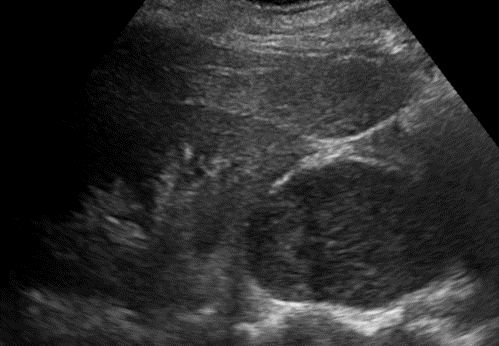
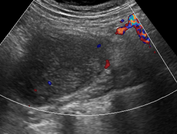
- Well-defined lesion, with internal echogenicity in the right paravertebral region, inferior to the right kidney, likely adjacent to or within the right psoas major muscle.
- Lesion demonstrates no internal vascularity.
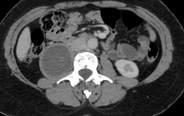
Contrast enhanced CT
- Well-defined hypo-enhancing lesion arising from the right L2-L3 neural foramina and extending into the right paravertebral region, insinuating into the right psoas major muscle, causing widening of the right L2-L3 neural foramina. However, no evidence of aggressive bone destruction.
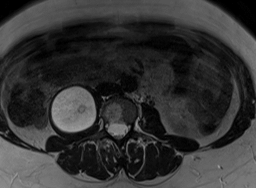
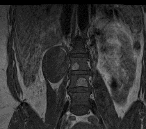
- Well-defined, predominantly T2 hyperintense lesion, arising from the right L2-L3 neural foramina and extending into the right paravertebral region, insinuating into the right psoas major muscle.
- The lesion demonstrates predominantly homogenous T2/STIR hyperintensity with small circular T2 hypointense areas interspersed within – fascicular sign.
Fascicular sign
- Multiple ring?like structures, which appear as hypointense foci within the hyperintense area on T2W images in neurogenic lesions -possibly reflecting the fascicular bundles.
- This sign is often seen in benign nerve sheath tumors
- The lesion demonstrates T1 hypointensity with T1 hyperintense crescentic areas in the upper and lower poles – suggestive of split fat sign, representing the intermuscular location of the lesion.
Split fat sign
- The presence of fat at the upper and lower poles of a lesion on T1W images -is suggestive of the intermuscular plane location of the lesion.
- Most commonly seen in neurogenic tumors, although it is not specific to them.
- A feature of benignity, as malignant lesions tend to be more infiltrative in nature, resulting in the obliteration of fat at the ends of the lesion.
- No evidence of restricted diffusion.

DIAGNOSIS:
- Intramuscular neurogenic tumor of benign etiology.
DISCUSSION:
- Peripheral nerve sheath tumors account for nearly 12% of the benign and 7?8% of the malignant soft tissue neoplasms.
- Neurofibromas and schwannomas constitute the benign category, while the malignant peripheral nerve sheath tumor comes under the malignant category.
- On MRI – fusiform lesions with low to intermediate signal intensity on T1W images and high?signal intensity on T2W images.
Differentiating Neurofibroma and Schwannoma:
- Both present as fusiform enlargement of the nerve, with the tapered ends of the lesion toward the parent nerve. Schwannomas are located eccentrically in relation to the nerve. Neurofibroma are central and inseparable from the parent nerve.
- T2 hyperintense rim – A thin rim of T2 hyperintensity has been observed more commonly in cases of schwannomas, as compared to neurofibromas
- Target sign – A central area of hypointensity with a peripheral hyperintensity on axial T2W images. Most often seen with neurofibromas. Postcontrast, there is central enhancement with peripheral hypointensity seen in these lesions.
- Intra?tumoral cystic degeneration is a more common feature of schwannomas, as compared to neurofibromas.
Benign vs malignant Schwannoma:
- Benign lesions usually < 5 cm and an increase in the size of these lesions is highly suggestive of a malignant transformation.
- Split fat sign – a feature of benignity, as malignant lesions tend to be more infiltrative in nature, resulting in the obliteration of fat at the ends of the lesion.
- Presence of perilesional edema adjacent to the primary lesion -suggestive of perilesional infiltration or edema
- The presence of a target sign in a malignant peripheral nerve sheath tumor is indicative of benign tissue within the lesion. The absence of this sign in a lesion is indicative of a malignant transformation of the primary benign lesion.
- Malignant lesions tend to be more heterogeneous on T1W and T2W images, secondary to the necrotic and hemorrhagic areas within.
REFERENCES
- Kakkar C, Shetty CM, Koteshwara P, Bajpai S. Telltale signs of peripheral neurogenic tumors on magnetic resonance imaging. Indian Journal of Radiology and Imaging. 2015 Oct;25(04):453-8.
Dr. Rashmi Jayakar Poojary
Radiology resident
Manipal Hospital, Yeshwanthpur, Bengaluru.
Dr. Anita Nagadi
Senior Consultant Radiologist
Manipal Hospital, Yeshwanthpur, Bengaluru
