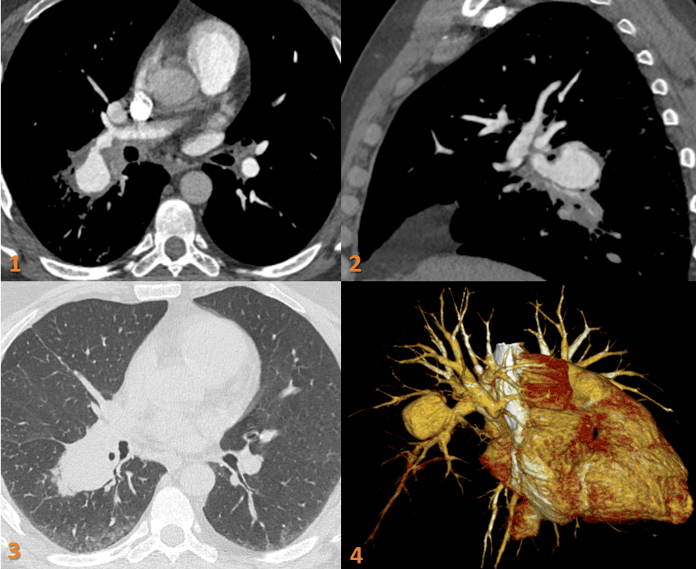
A 43-year gentleman with recurrent hemoptysis, recurrent genital and oral ulcers since 5 years.
- Fig 1 & 2. Axial and Sagittal contrast CT Pulmonary angiogram images demonstrate a large saccular aneurysm arising from the superior segmental artery of right lower lobar pulmonary artery with diffuse wall thickening – probable eccentric thrombus. There is compression of distal lower lobe segmental pulmonary arteries by the aneurysm (arrow).
- Fig 3. Axial CT image Lung window: Perifocal ground glass opacity along the saccular aneurysm in the right lower lobe.
- Fig 4. VR image of pulmonary artery aneurysm.
Post coiling of pulmonary artery aneurysm.
DIAGNOSIS:
Right pulmonary artery aneurysm with partial eccentric thrombus.
DISCUSSION:
Behçet disease is a multisystemic and chronic relapsing inflammatory disorder characterized by the histopathologic finding of nonspecific vasculitis involving various sized vessels in multiple organs.
Key imaging manifestations are:
- Pulmonary thromboembolism.
- Pulmonary artery aneurysm.
- Sinus of Valsalva aneurysm.
- SVC syndrome.
- Intracardiac thrombosis.
- Mediastinal fibrosis.
- Intestinal ulceration (and inflammatory changes).
- Budd-Chiari syndrome.
- Cerebral venous thrombosis.
- Identification of a vascular lesion is important because it seriously affects a patient’s prognosis. The leading cause of sudden death in patients with Behçet disease is rupture of a large aortic or arterial aneurysm
- To improve the detection of these various radiologic findings, examinations should be well-selected and optimized in patients with Behçet disease.
REFERENCES:
- Mehdipoor G, Davatchi F, Ghoreishian H, Shabestari AA. Imaging manifestations of Behcet’s disease: key considerations and major features. European journal of radiology. 2018 Jan 1;98:214-25.
- Chae EJ, Do KH, Seo JB, Park SH, Kang JW, Jang YM, Lee JS, Song JW, Song KS, Lee JH, Kim AY. Radiologic and clinical findings of Behçet disease: comprehensive review of multisystemic involvement. Radiographics. 2008 Sep;28(5):e31.
Dr. Deepali Saxena,
DNB, Fellowship Cardiothoracic Imaging (USA)
Lead Cardiothoracic Imaging
Manipal Hospitals Radiology Group.
Unit coordinator, Whitefield
Dr. Vivek Jirankali,
MD
Senior resident and cross-sectional fellow
Manipal Hospitals Radiology Group.

