39-year-old male with increased frequency of stools, intermittent bilateral iliac region pain, progressive abdominal distension, and weight loss of 12kgs in the last 4 months
-
39-year-old male with increased frequency of stools, intermittent bilateral iliac region pain, progressive abdominal distension, and weight loss of 12kgs in the last 4 months
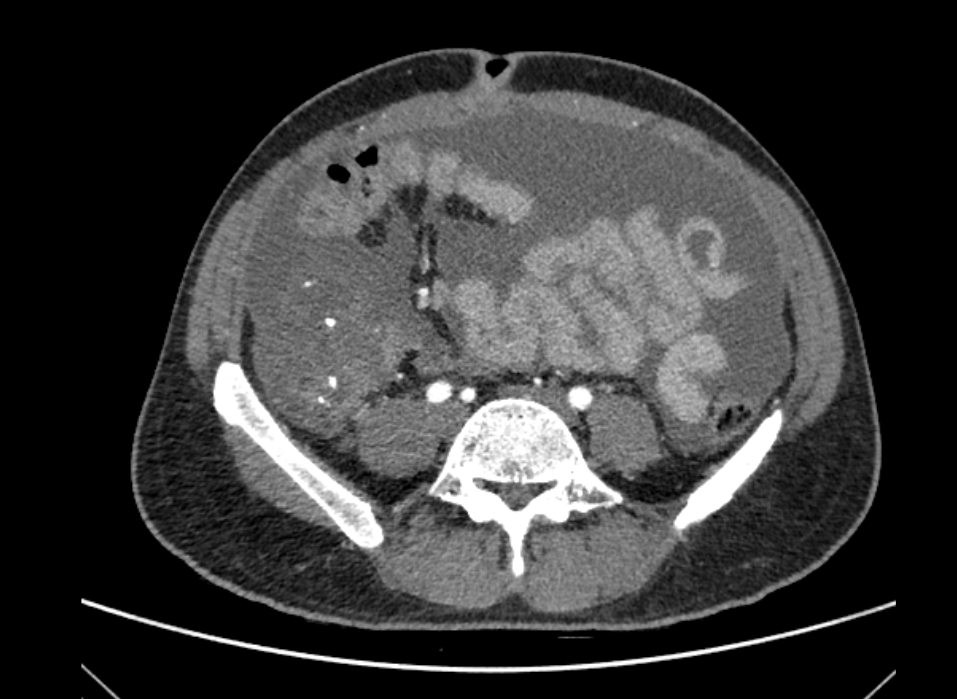

- Ill-defined, hypodense lesion in the right iliac fossa, with coarse linear and curvilinear calcifications. There is loss of fat plane with the psoas major posteriorly.
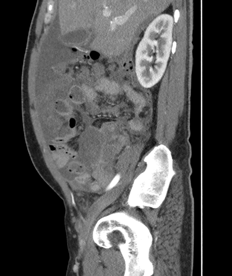
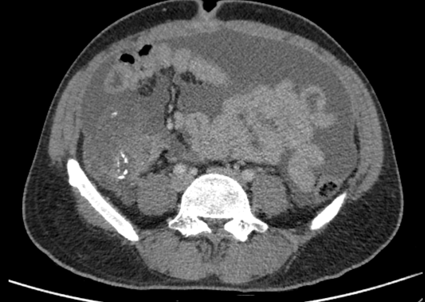
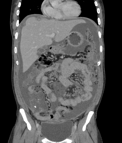
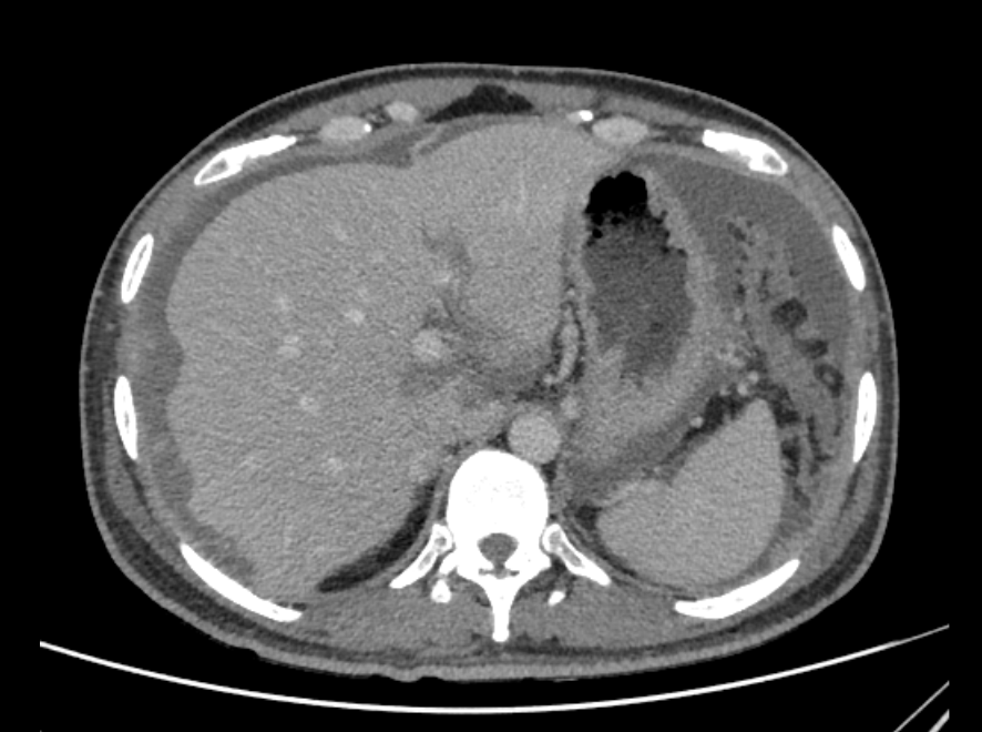
- Low density fluid in the peritoneal cavity, with scalloped margins along the surface of the right lobe of the liver – suggestive of pseudomyxoma peritonei.
- Surface deposits along the underside of the left liver lobe.Diffuse omental nodularity present.
RADIOLOGICAL DIAGNOSIS
- Ill-defined, hypodense lesion in the right iliac fossa, with coarse linear and curvilinear calcifications.
- Low density fluid in the peritoneal cavity, with scalloped margins along the surface of the right lobe of the liver – suggestive of pseudomyxoma peritonei.
- Surface deposits noted along the underside of the left liver lobe.
- Extensive omental deposits.
- Mild left pleural effusion.
Imaging features are suggestive of appendiceal mucinous adenocarcinoma.
Discussion:
- Mucinous neoplasms comprise approximately 70% of the epithelial tumors of the appendix. The characteristic expansile spread of these tumors, as opposed to classic infiltrative invasion.
- Appendiceal mucinous neoplasms are classified into four types: Adenoma, LAMN (low-grade appendiceal mucinous neoplasm), HAMN (high-grade appendiceal mucinous neoplasm), and mucinous adenocarcinoma.
- The classic radiologic manifestation of an appendiceal mucinous tumor is a mucocele.
- In addition, LAMN, HAMN, and mucinous adenocarcinoma may demonstrate extra-appendiceal spread of mucin, which may be confined to the peri-appendiceal region or disseminated as PMP (pseudomyxoma peritonei).
- Mural nodularity and irregular wall thickening are features that have been associated with malignant mucoceles due to adenocarcinoma.
- The shape, attenuation of internal content, maximal wall thickness, and presence of internal septa, wall calcifications, peri-appendiceal fat stranding, or intraperitoneal free fluid are not helpful in differentiating malignant from benign mucoceles.
- PMP is a clinical syndrome characterized by the presence of mucin and/or epithelial cells within the peritoneum and on the serosa of the abdominal and pelvic organs.
- The primary neoplasm causing PMP is almost always appendiceal in origin, with other less common sites including the ovary, colon, urachus, and pancreas.
- Imaging manifestations of PMP include mucinous ascites, peritoneal soft-tissue implants, omental caking, and involvement of the gastrointestinal tract and ovaries. Cross-sectional imaging demonstrates loculated peritoneal collections that can progress to large-volume ascites. The solid abdominal organs develop a scalloped appearance due to mass effect by tumoral implants, and parenchymal invasion can occur at the sites of serosal involvement. Linear or punctate calcifications of the mucinous deposits may be identified.
- The hollow viscera are displaced and distorted, and small-bowel obstruction due to tumor deposits is a common complication in advanced disease.
REFERENCE:
- Neoplasms of the appendix – Pictorial review with clinic-pathological correlation: Laura M et al – Radiographics.
- Pseudomyxoma from mucinous adenocarcinoma f the appendix extending from the retroperitoneum into the thigh – Beth V et al.
- Mucinous neoplasms of the appendix: a current comprehensive clinicopathologic and imaging review – Sree Harsha Tirumani et al.
Dr. Madhu Kumar S B
Senior Consultant Radiology
Manipal Hospital, Yeshwanthpur, Bengaluru.
Dr. Priyanka Rout
Radiology Resident
Manipal Hospital, Yeshwanthpur, Bengaluru.