A 36-year-old with history of multiple episodes of seizure
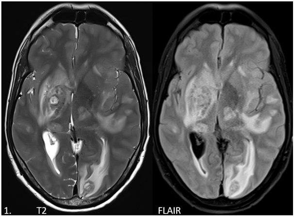
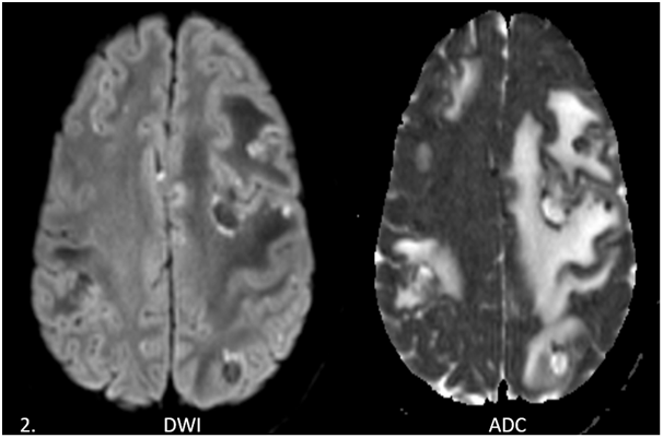
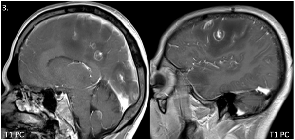
- Multifocal supra-tentorial and infra-tentorial, ring enhancing intra-axial lesions in deep grey matter and at grey-white matter junction in bilateral cerebral lobes.
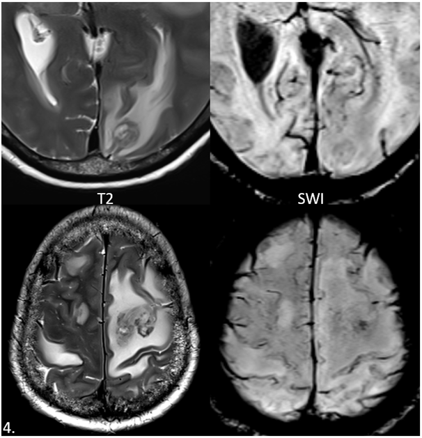
- The lesions demonstrate heterogenous T2 signal, T1 hypointense signal with peripheral restricted diffusion and patchy foci of susceptibility on SWI representing haemorrhages with moderate perilesional vasogenic edema.
- Most of these lesions demonstrates an eccentric nodule along the rim of an enhancing lesion – eccentric target sign.
DIAGNOSIS AND DISCUSSION
Neuro-toxoplasmosis.
DISCUSSION:
- Toxoplasmosis is caused by Toxoplasma gondii, an intracellular protozoan that is found worldwide.
- It is transmitted to humans primarily by ingestion of cysts in undercooked pork or lamb or contaminated vegetables or through direct contact with cat faeces.
- Our patient (PLHIV) on further work up was found to have a CD4 count of 24 cells/µL and Serum Toxoplasma IgG – 11.5 (elevated).
- CD4 count of <100 cells/µL, increases the risk of opportunistic infections like toxoplasmosis, candida, histoplasmosis etc….
- Typically, the lesions are seen in basal ganglia, thalami and corticomedullary junction.
- Presence of eccentric nodule in ring enhancing lesions and intralesional susceptibility focus on SWI, is almost considered pathognomonic imaging signs for CNS toxoplasmosis.
- On PET-CT/Thallium SPECT minimal or no uptake helps to differentiate from CNS lymphoma.
- Differential diagnosis includes CNS Lymphoma, Metastasis and other CNS infections like TB, NCC etc.
References:
- Gregory Tse Lee, Fernando Antelo, and Anton A. Mlikotic. Cerebral toxoplasmosis. RadioGraphics 2009 29:4, 1200-1205
- Kumar GG, Mahadevan A, Guruprasad AS, et al. Eccentric target sign in cerebral toxoplasmosis: neuropathological correlate to the imaging feature. J Magn Reson Imaging. 2010;31(6):1469-1472. doi:10.1002/jmri.22192
- Benson JC, Cervantes G, Baron TR, et al. Imaging features of neurotoxoplasmosis: A multiparametric approach, with emphasis on susceptibility-weighted imaging. Eur J Radiol Open. 2018;5:45-51. Published 2018 Mar 17. doi:10.1016/j.ejro.2018.03.004
Dr.Harsha C Chadaga
DMRD,DNB,PDCC,EDiNR
Senior Consultant and Head of Radiology
Manipal Hospitals Radiology Group.
Dr. Diwakar C
Radiology resident
Manipal Hospitals Referral hospital


