

A 35 year old female with complaint of lump at posterior aspect of tongue
A 35-year-old female with a complaint of a lump at the posterior aspect of her tongue.
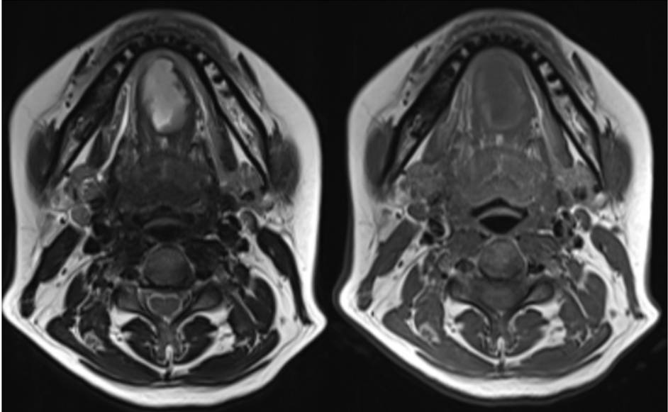
- Axial T2w and T1w sequences demonstrate an irregular T2 hyperintense, T1 hypointense lesion with a T1 hyperintense rim in the midline floor of mouth/sublingual space, infiltrating the genioglossus muscle.
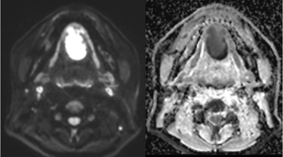
- Axial DWI and ADC demonstrate diffusion restriction.
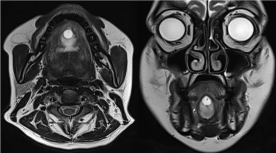
- Axial and coronal T2w sequences demonstrate a well circumscribed midline cyst in the anterosuperior aspect of the lesion. There is an eccentric T2 hypointense dot within the cyst. There is associated T2 hyperintense oedema with mild swelling in the anterior 2/3rds of the tongue.
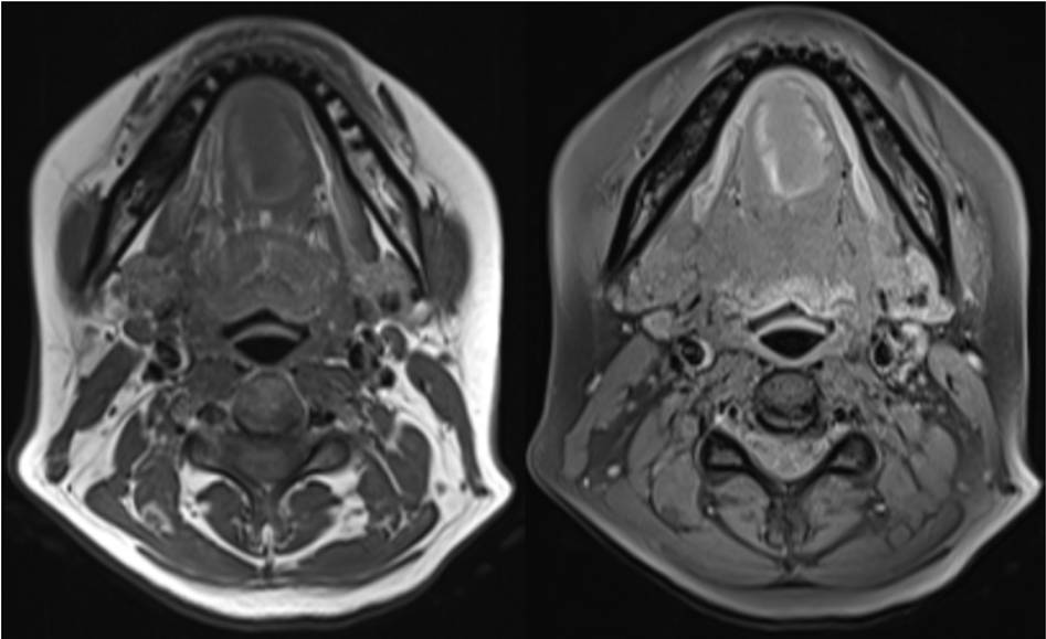
- Axial pre-contrast and post contrast T1w fat saturated images demonstrate the irregular peripheral enhancement of the lesion.
Diagnosis:
Sublingual cysticercosis with abscess formation.
Discussion:
Cysticercosis of the oral cavity is rare with floor of mouth lesion being the rarest among all oral lesions.
Various appearances have been described:
- The commonest being that of a cyst with a scolex and a surrounding inflammatory mass or an abscess. This may occur due to chronic leakage of cyst fluid or secondary bacterial infection.
- Another appearance is that of a large oedematous fluid collection with a scolex situated eccentrically.
- Sometimes, the scolex may escape and not be seen and the collection may be indistinguishable from an abscess.
- Finally, a rare appearance is that of a calcified cyst.
MR imaging is particularly useful in disseminated cases in detecting cysticerci and their complications.
- Cysticerci appear as well-defined oval or rounded T2 hyperintense and T1 hypointense lesions, often aligned parallel to the long axis of the muscle fibers (in case of muscular cysticerci).
- On fat suppressed, post-gadolinium T1 images, mild perilesional enhancement of the cyst may be seen.
- Abscesses appear as heterogenous T2 hyperintense and T1 hypointense lesions which often reveal a T2 hypointense rim due to presence of free radicals.
Differential diagnosis:
Ruptured retention cyst/mucocele of minor salivary gland with abscess. In such cases no eccentric dot (scolex) will be seen.
References:
- Muthyala S, Krishna KV, Kishan TV, Bhuvana NS, Moorthy RS. Masseteric cysticercosis with abscess formation: A diagnostic dilemma. Medical Journal, Armed Forces India. 2015 Jul;71(Suppl 1):S148.
- Manoj P, Naveen S, Vineeta S, Devendra K, Mohan K. Floor of mouth cysticercosis. World Journal of Medical and Surgical Case Reports. 2012 Jul 30;1(1).
Dr. Vivek Jirankali MD,
Senior Resident and Cross-Sectional Fellow,
Manipal Hospitals Radiology Group.
Dr. Anita Nagadi,
MD, MRCPCH, FRCR, CCT (UK)
Lead Head & Neck, Oncology
Senior Consultant Radiologist
Manipal Hospitals Radiology Group.
