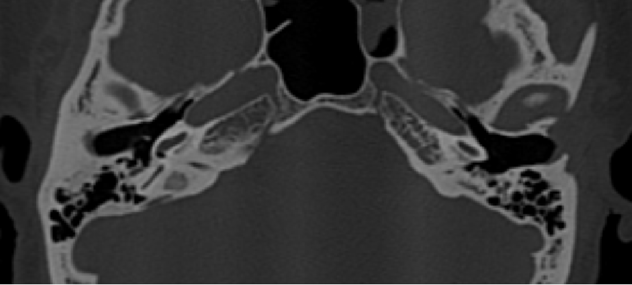
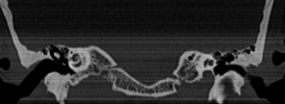
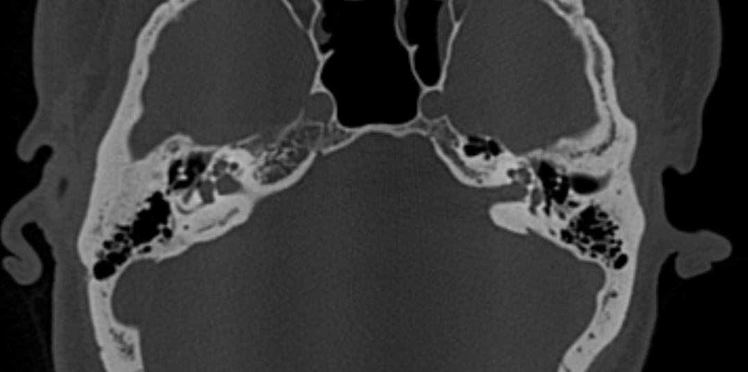
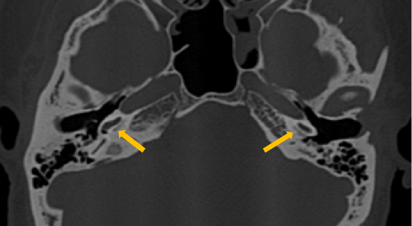
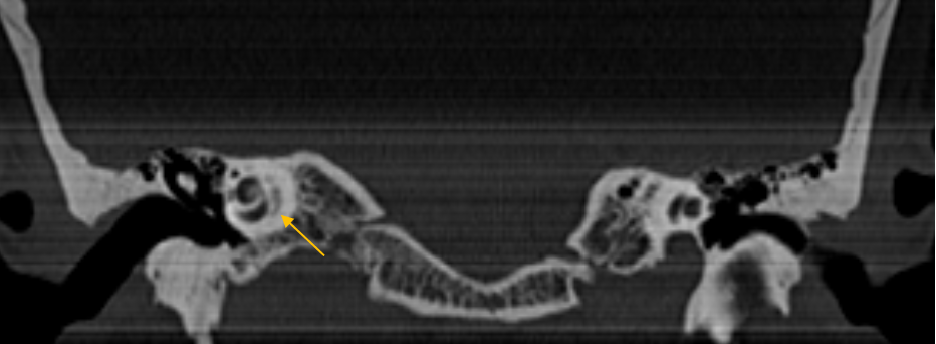
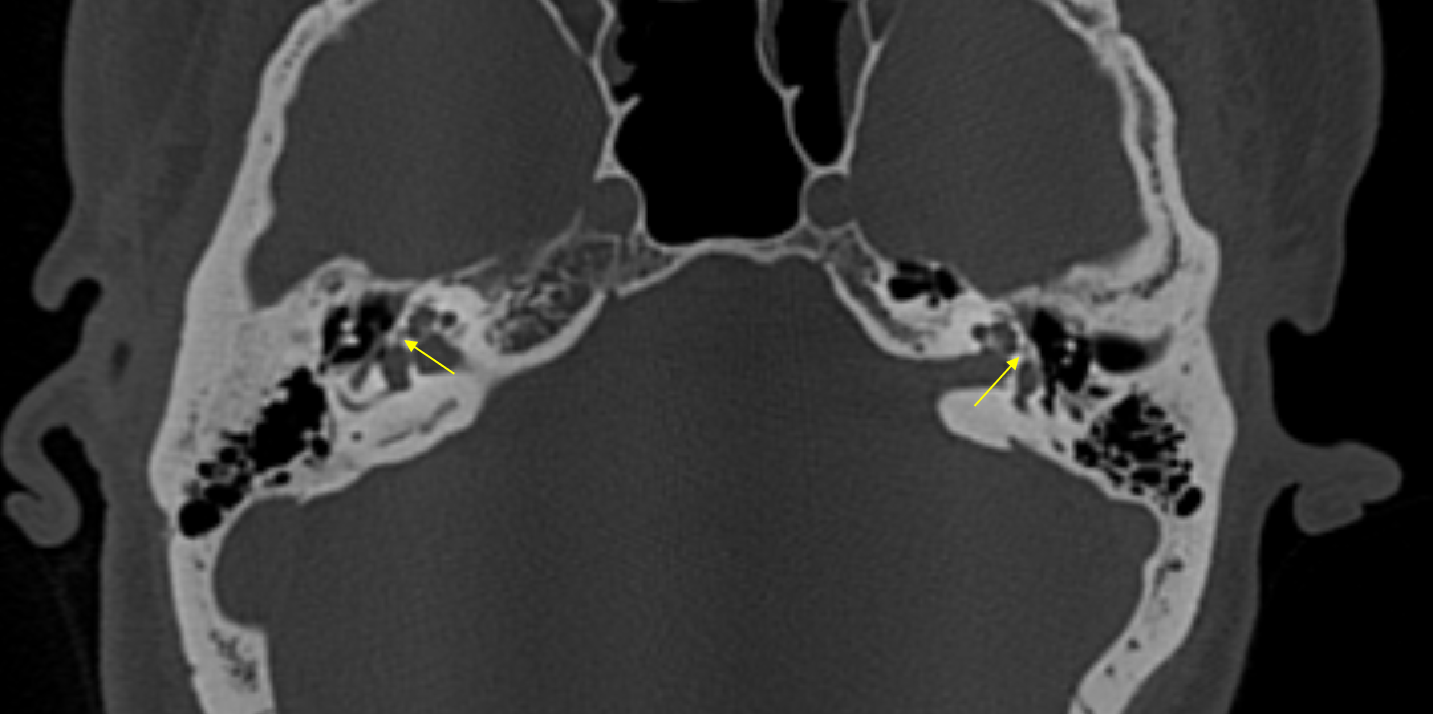
A 33 year old, male with bilateral mixed hearing loss.
- Lucencies around bilateral cochlea, representing demineralization (yellow arrow) – double ring sign.
- Foci of lucencies involving the fissula ante fenestrum on both sides.
Diagnosis:
- Bilateral fenestral and retrofenestral otosclerosis.
Discussion:
- Otosclerosis is a misnomer, as it is characterised by lucent rather than sclerotic bony changes and hence more appropriate term is otospongiosis.
- Female predilection is present with a F:M ratio of ~2:1.
- Most commonly presents with hearing loss, often conductive, but can also be sensorineural or mixed and is frequently bilateral.
- Hearing loss may be exacerbated by pregnancy.
- Pure-tone audiometry shows characteristic decrease in bone conduction at 2000 Hz (Carhart notch).
2 Subtypes:
- Fenestral (stapedial)
- anterior to the oval window, involving a small cleft known as the fissula ante fenestram
- conductive hearing loss due to stapes thickening and fixation
- Retrofenestral(cochlear)
- demineralisation of the cochlear capsule
- hearing loss is often sensorineural
Grading of otosclerosis (Symons and Fanning)
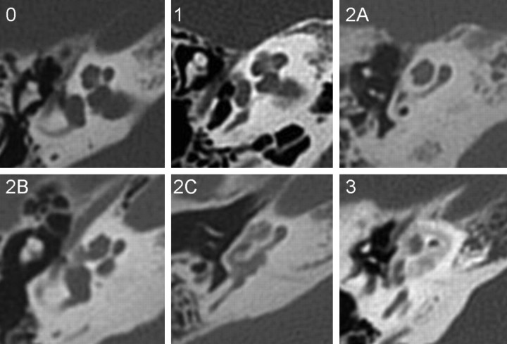
- Grade 0: normal.
- Grade 1: small lucent lesion at the fissula ante fenestram.
- Grade 2A: sclerosis and narrowing of the basal turn.
- Grade 2B: lucent lesion extending from the fissula ante fenestram to the middle turn of the cochlea.
- Grade 2C: patchy lucency around the lateral aspect of basal, middle, and apical turns of the cochlea, the medial aspect of the cochlea appears spared.
- Grade 3: severe, confluent lucency around the cochlea.
References:
- Lee TC, Aviv RI, Chen JM, Nedzelski JM, Fox AJ, Symons SP. CT grading of otosclerosis. American journal of neuroradiology. 2009 Aug 1;30(7):1435-9.
- Purohit B, Hermans R, Op de Beeck K. Imaging in otosclerosis: a pictorial review. Insights into imaging. 2014 Apr;5:245-52.
Dr Anita Nagadi
Senior Consultant Radiologist
Manipal Hospital, Yeshwanthpur, Bengaluru
Dr Rashmi Jayakar Poojary
Radiology Resident
Manipal Hospital, Yeshwanthpur, Bengaluru

