
A 26-year gentleman with reduced left sided vision, proptosis and headache
A 26-year gentleman with reduced left-sided vision, proptosis, and headache.
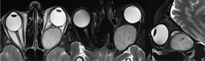
- Axial T2/STIR and sagittal STIR images demonstrate a well-defined oval shaped mass lesion in intra conal space of left orbit just behind the globe significantly compressing and displacing the intraorbital segment of left optic nerve superomedially with significant thinning and T2 hyperintensity.
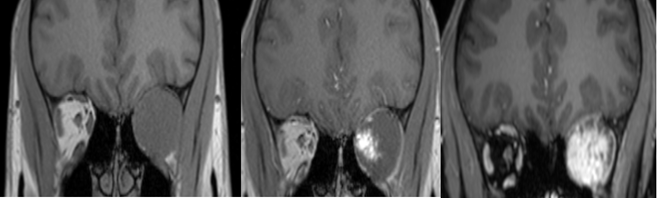
- Axial T1w precontrast, immediate post-contrast and delayed post-contrast images demonstrate focal and patchy enhancement on the immediate postcontrast images with gradual filling-in and heterogeneous enhancement on delayed post-contrast images.
DIAGNOSIS:
Cavernous hemangioma.
DISCUSSION:
- Cavernous hemangioma is the most common vascular lesion of orbit in adults.
- Cavernous malformations occur most often in women (60%–70%) between the ages of 18 and 72 years (mean age, 43– 48 years), slowly and progressively enlarge, and do not involute. Most often occur in the lateral aspect of the retrobulbar intraconal space, are rarely intramuscular and uncommonly involve the orbital apex.
- They extend intracranially through the superior orbital fissure. Bone remodeling is not uncommon, and intralesional calcification occurs occasionally.
- At MR imaging, the signal in cavernous malformations usually appears isointense to that of muscle on T1-weighted images and hyperintense to that of muscle on T2-weighted images. Internal septa are visible within larger lesions. Cavernous malformations demonstrate progressive accumulation of contrast material on late phase dynamic images and delayed images.
- Conventional angiography with a prolonged injection may demonstrate delayed contrast material pooling, a feature that permits the differentiation of cavernous malformations from other vascular lesions.
DIFFERENTIAL DIAGNOSIS:
ORBITAL SCHWANNOMA:
- Schwannomas are typically extraconal and located at the superior orbit, owing to their frequent origin from the frontal branch of the ophthalmic nerve.
- Cystic changes are more frequent with heterogeneously hyperintense signal on T2-weighhted images.
- In the early phase, all the hemangiomas show enhancement from one point or portion with gradual filling as compared to schwannomas which show enhancement from a wide area on early phase. Hence, hemangioma and schwannoma of the orbit can be differentiated by the contrast-enhancement spread pattern on dynamic MRI.
REFERENCES:
- Smoker WR, Gentry LR, Yee NK, Reede DL, Nerad JA. Vascular lesions of the orbit: more than meets the eye. Radiographics. 2008 Jan;28(1):185-204.
- Tanaka A, Mihara F, Yoshiura T, Togao O, Kuwabara Y, Natori Y, Sasaki T, Honda H. Differentiation of cavernous hemangioma from schwannoma of the orbit: a dynamic MRI study. American Journal of Roentgenology. 2004 Dec;183(6):1799-804.
Dr. Sriram S Patwari MD, PDCC.
Consultant Radiologist and Co-lead Neuroradiology
Manipal Hospitals Radiology Group.
Dr. Vivek J. MD
Senior Resident and Cross-sectional fellow
Manipal Hospitals Radiology Group.