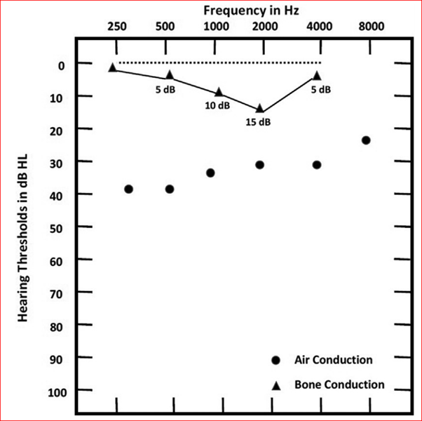
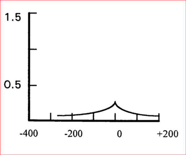
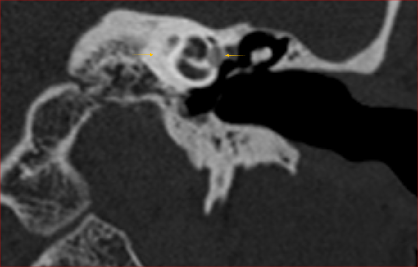
A 25 years old man, presented with complaints of left sided hearing loss and tinnitus. On otoscopy, bilateral tympanic membranes were intact.
A 25 years old man, presented with complaints of left sided hearing loss and tinnitus.
On otoscopy, bilateral tympanic membranes were intact.
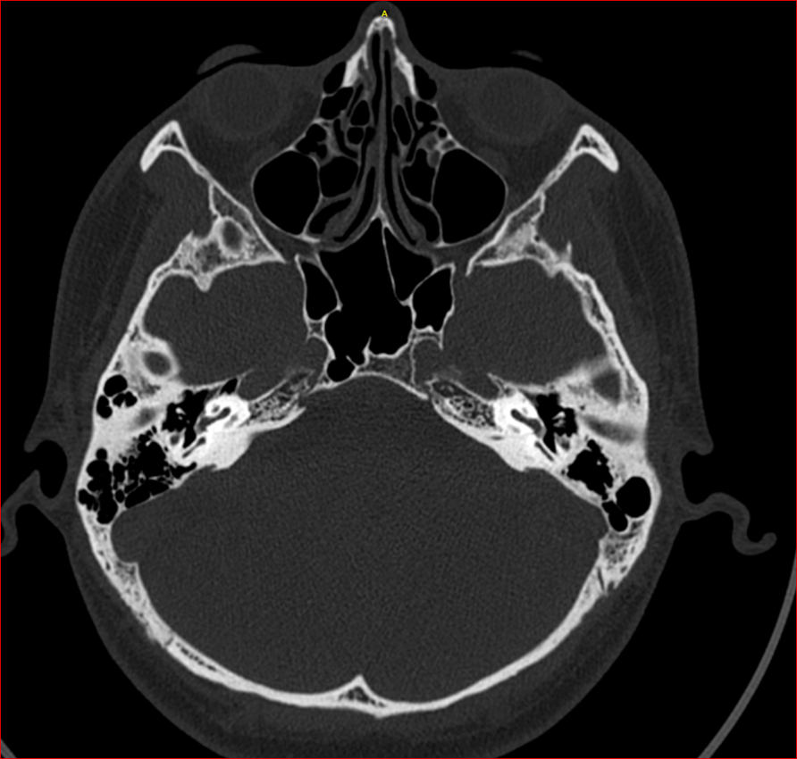
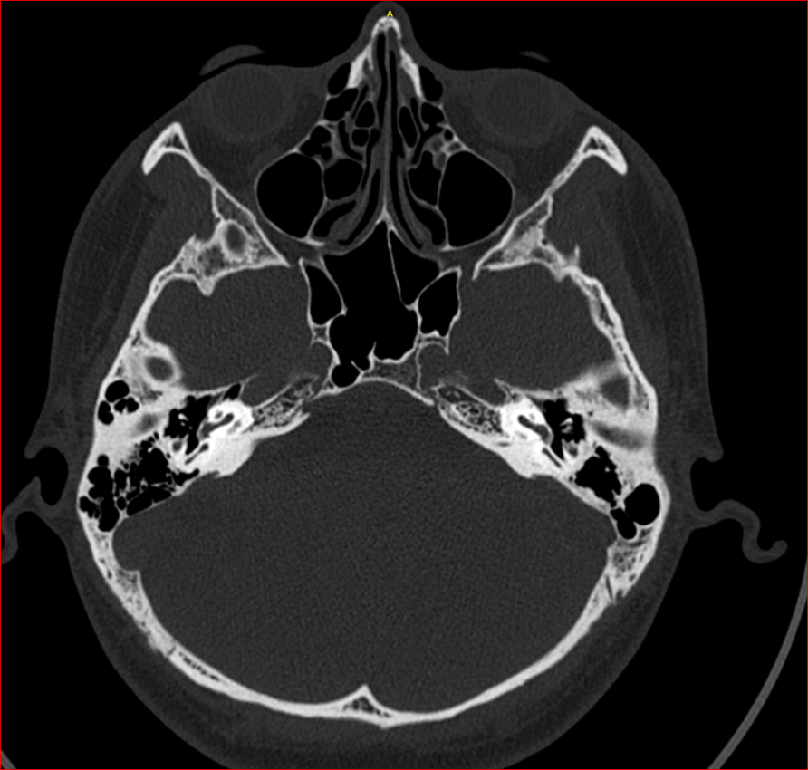
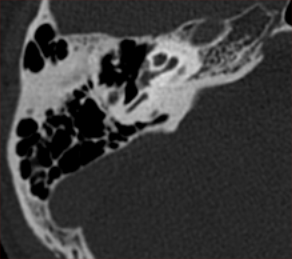
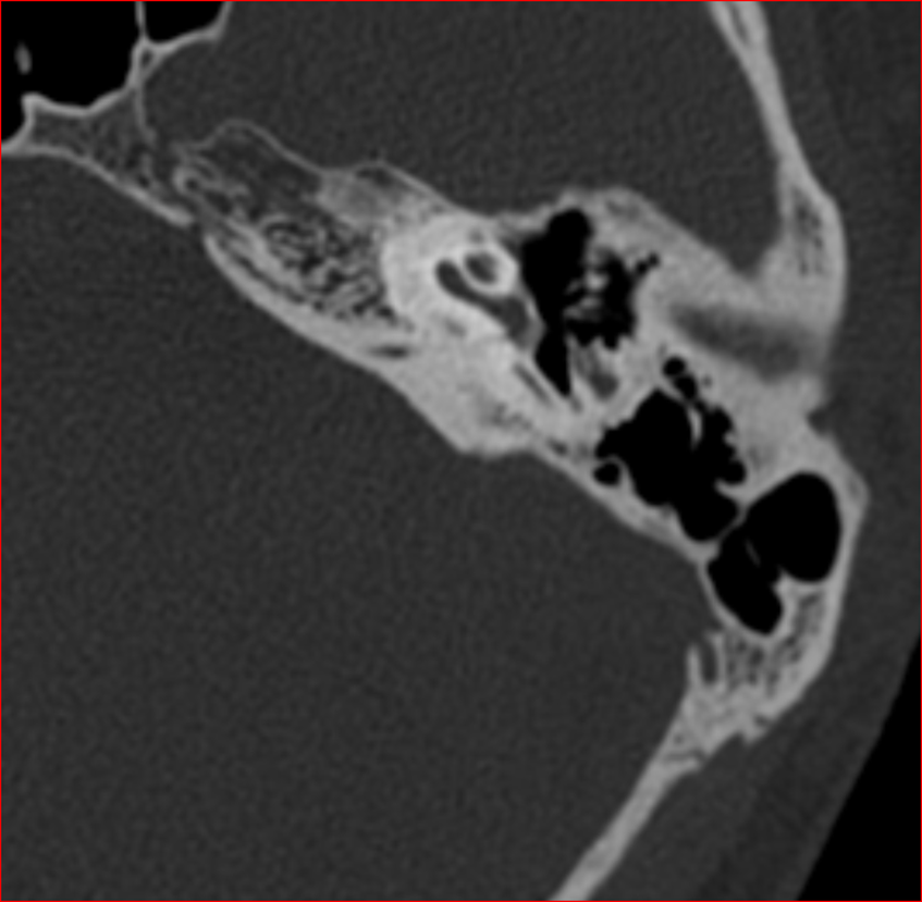
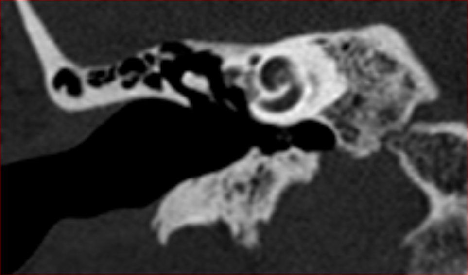
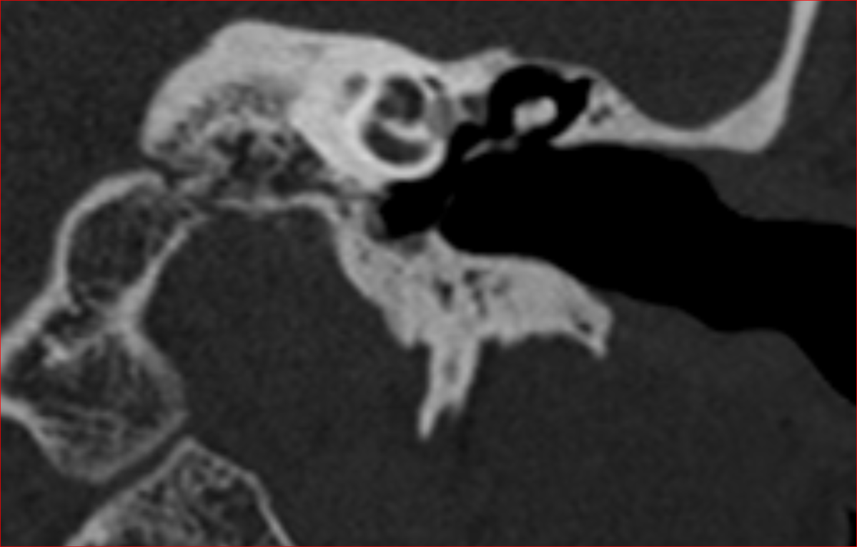
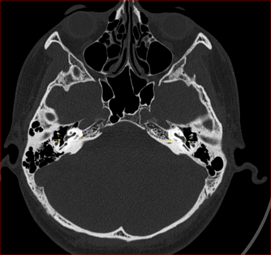
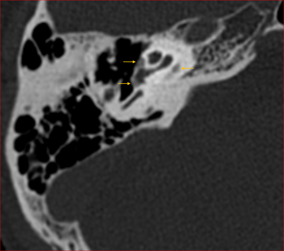
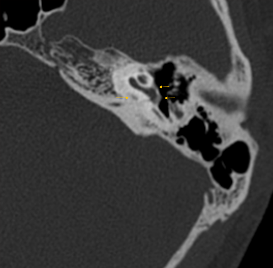
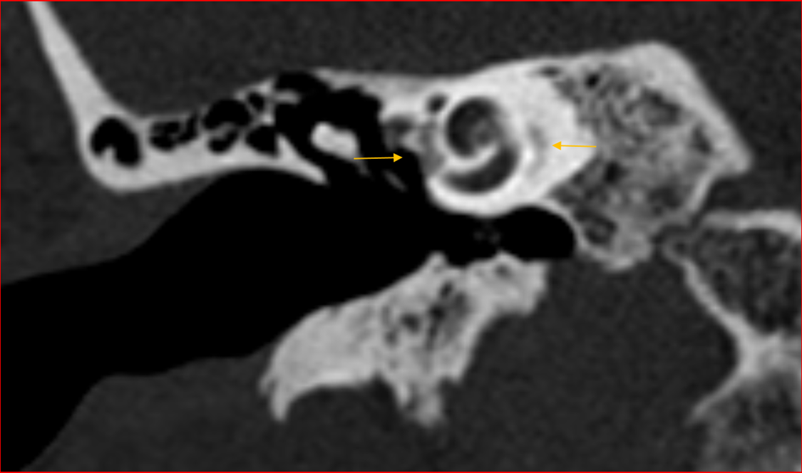
Axial and coronal HRCT images of the temporal bones demonstrate large foci of demineralization in the region of the fissula ante fenestram, cochlear promontory, round window niche and retro cochlear regions bilaterally.
Diagnosis:
Fenestral and retro fenestral otosclerosis.
Discussion:
Otosclerosis is a primary osteodystrophy of the otic capsule.There are two subtypes:
- Fenestral? (stapedial): Most common
involves the?oval window?and the?stapes?footplate ?conductive hearing loss
- Retro fenestral? (cochlear):
cochlear?involvement with demineralization of the cochlear capsule sensorineural hearing loss
Key diagnostic features:
- Most common location to be involved fissula ante fenestram
Findings depend on the phase of the disease:
A. Oto spongiotic phase:
- There is demineralization of the endochondral layer of the bony labyrinth and formation of spongy bone
- Manifests as decreased attenuation (lucency) within the normally homogeneously dense border of the otic capsule
B. Oto sclerotic phase
- The region increases in attenuation
- In severe cases, the oval window and/or round window are completely filled in by a dense bony plate
Treatment
- If the patient has mild hearing loss no treatment is needed.
- In patients with moderate conductive loss either hearing aid or operative treatment by way of small fenestra stapedotomy can be considered.
- In small fenestra stapedotomy the fixed footplate is bypassed by a prosthesis connecting the long process of incus with footplate of stapes.

Dr. Rosmi Hassan K
Radiology Resident
Manipal Hospitals Radiology Group
Dr. Anita Nagadi
MD, MRCPCH, FRCR, CCT
Senior Consultant Radiology
Manipal Hospitals Radiology Group

