

A 25- years-old male presented with swelling at wrist and no history of trauma
- 25y, Male, presenting with.
- No H/o trauma.
FINDINGS
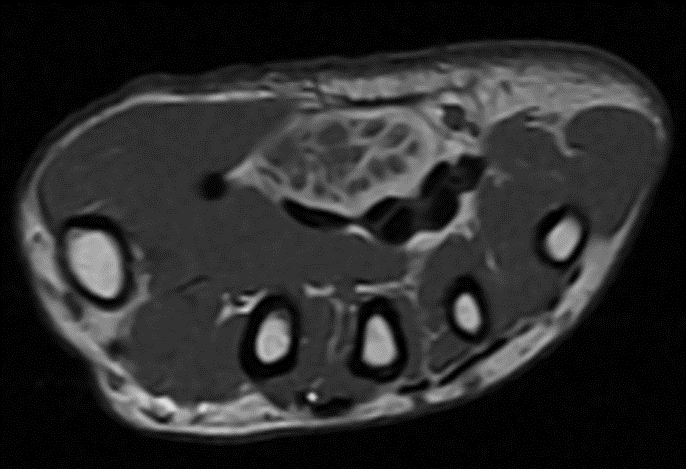
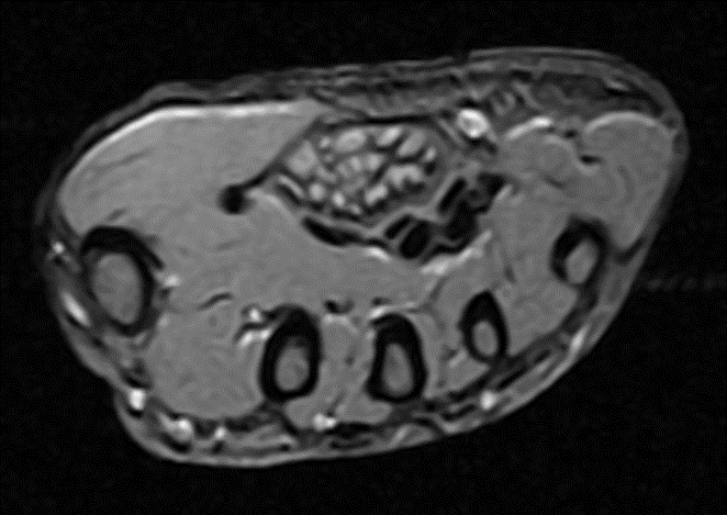
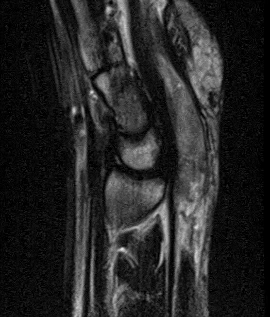
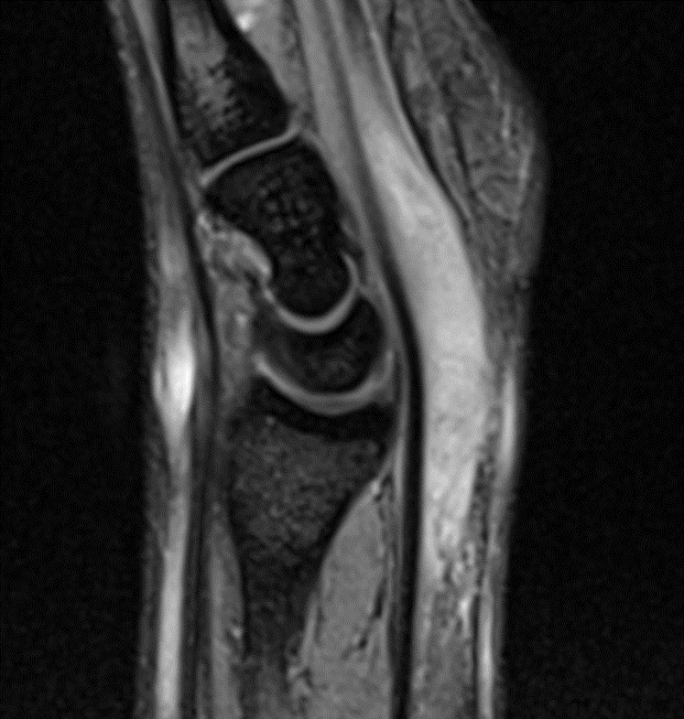
- Short segment gross thickening of the median nerve (blue arrow) seen below the flexor retinaculum (Flexor retinaculum).
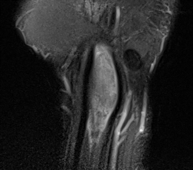
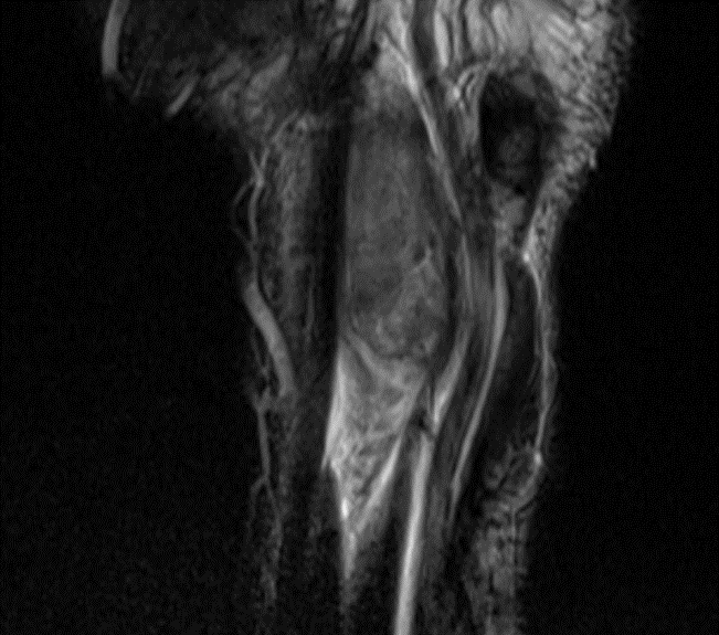
- There is lipomatous tissue interdigitating between the prominent fascicles giving coaxial cable appearance on axial and sphagetti like appearance on coronal sequences.
- Adjacent flexor tendons (orange arrow) and rest of neurovascular structures are normal.
Diagnosis: Fibro lipohamartoma of median nerve
DISCUSSION:
- Is a benign neoplasm of nerves, resulting from anomalous growth of fibroadipose tissue contained by epineurium resulting in expansion of the nerve with separation of nerve fascicles.
- When enlargement of the nerve results in visible enlargement and deformity of the body, most commonly the hand, then the term macro dystrophia lipomatosis is often used, although the conditions are essentially synonymous.
Epidemiology
- Lipomatosis of nerve is most commonly encountered in children before the age of 10 years of age.
Clinical presentation
- This condition either presents as a slowly growing asymptomatic mass, or with pain and paresthesia (most common) particularly if associated with nerve compression or entrapment.
- For example, carpal tunnel syndrome is common when the median nerve is involved in the carpal region.
- When digital branches of nerves are involved this can result in macrodactyly of the involved digit.
Pathology
- Lipomatosis of nerve is characterized by hamartomatous overgrowth of mature fat and fibroblasts within the epineurium resulting in marked expansion of the nerve.
- Other than case reports with associated proteus syndrome and Klippel-Trénaunay syndrome, there are no known syndromic associations and the condition appears sporadic.
Location
- Lipomatosis of nerve is almost exclusively seen in the limbs, most commonly in the upper limb.
- Only rarely is bilateral or found elsewhere.
- upper limb: 74%.
- median nerve: >60%
- ulnar nerve: 7%
- lower limb: 17%
- plantar nerve: 11%
Classification
The WHO classification of tumors of soft tissue categorizes this entity under adipocytic tumors.
Radiographic features
Ultrasound
A characteristic hypoechoic coaxial cabling encased by an echogenic substratum may be seen.
MRI
MRI features are often pathognomonic and typically shows a coaxial cable-like appearance on axial images and a spaghetti-like appearance on coronal images. A pseudo-union-bulb appearance may be present.
- T1:the neural bundles are hyperintense to muscle and the surrounding substratum is isointense to muscle.
- T2:fat components are high signal and fibrous components are low signal.
Treatment and prognosis
Lipomatosis of nerve is benign and therefore treatment is for symptoms but needs to be balanced with preservation of nerve function as it is difficult to resect the lesion without also resecting/impinging the affected nerve.
REFERENCES
- Silverman TA, Enzinger FM. Fibrolipomatous hamartoma of nerve. A clinicopathologic analysis of 26 cases. Am. J. Surg. Pathol. 1985;9 (1): 7-14. – Pubmed citation.
- Jain TP, Srivastava DN, Mittal R et-al. Fibrolipomatous hamartoma of the median nerve. Australas Radiol. 2007;51 Spec No. : B98-B100. doi:10.1111/j.1440-1673.2007.01790.x – Pubmed citation
- Toms AP, Anastakis D, Bleakney RR, et al. Lipofibromatous hamartoma of the upper extremity: a review of the radiologic findings for 15 patients. AJR Am J Roentgenol. 2006;186 (3): 805 11. doi:10.2214/AJR.04.1717 – Pubmed citation.
- Murphey MD, Carroll JF, Flemming DJ, et al. From the archives of the AFIP: benign musculoskeletal lipomatous lesions. Radiographics. 24 (5): 1433-66. doi:10.1148/rg.245045120 – Pubmed citation
- Mason ML. Presentation of cases. Proceedings of the American Society of Surgery of the Hand. J Bone Joint Surg Am. 1953 Jan 1;35A(1):273–275.
Dr. Praveen Wali
Consultant Radiologist.
Manipal Hospital Radiology Group (MHRG)
Manipal Hospital, Bengaluru.
Dr. Srinivas P
Fellow in Radiology
Manipal Hospital Radiology Group (MHRG)
Manipal Hospital, Bengaluru.
