
70 Year Female Patient who is K/c/o Rheumatoid Arthritis with CKD and Type 2 DM with past H/o pericardial effusion.
History
70/Female patient who is K/c/o Rheumatoid Arthritis with CKD and Type 2 DM with past H/o pericardial effusion.
Now presented with echogenic mass in pericardium on echocardiography
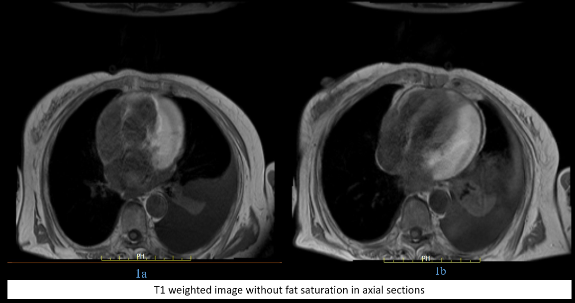
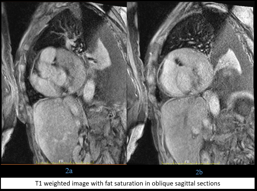
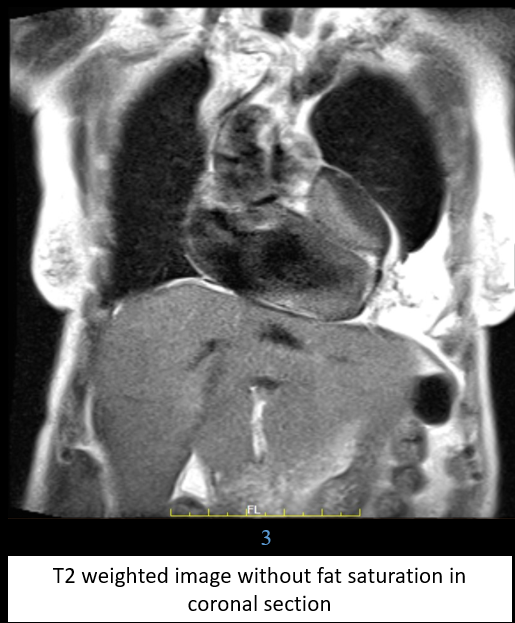
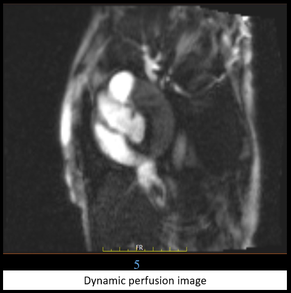
Blue arrow – lesion: organized hematoma or organized pericardial effusion.
Orange arrow – thickened pericardium
Red arrow – normal myocardium
Green arrow – areas of calcification seen as blooming on MRI (CT correlated)
DIAGNOSIS AND DISCUSSION:
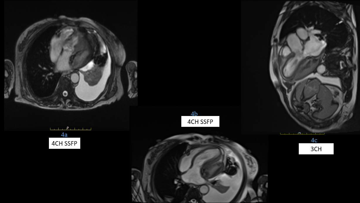
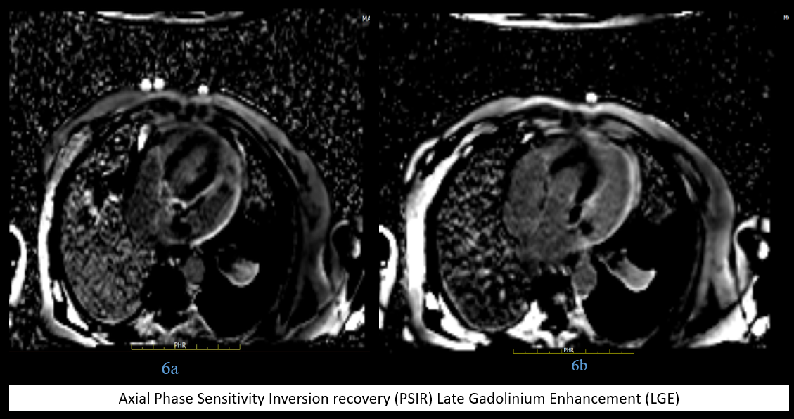
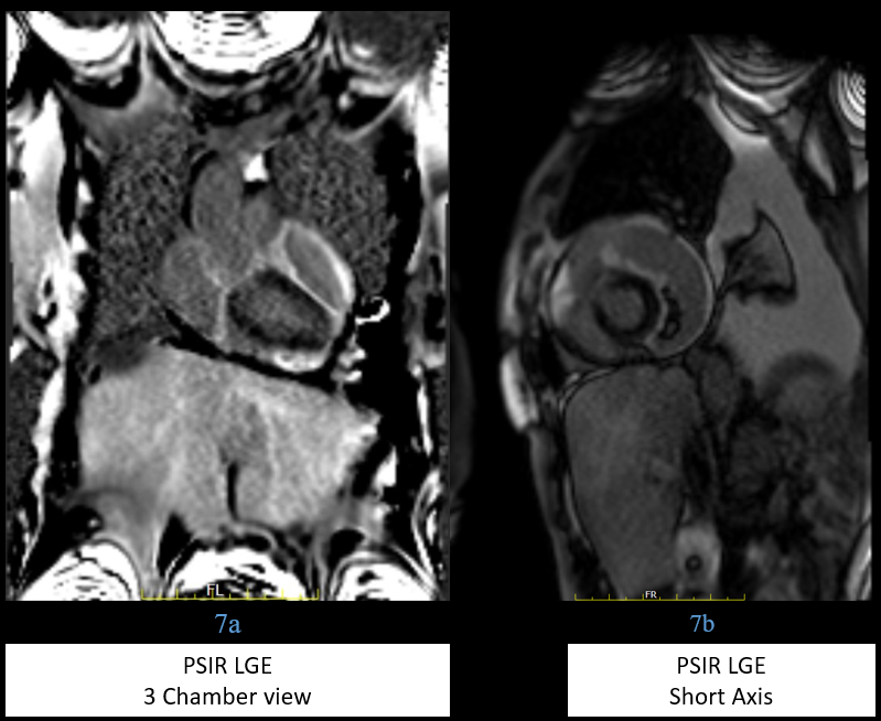
LESION ALONG LV WALL WITH PERICARDIAL CONSTRICTION WITH POSSIBLE DIFFERENTIALS
1. Organised pericardial hematoma.
2. Organised pericardial effusion.
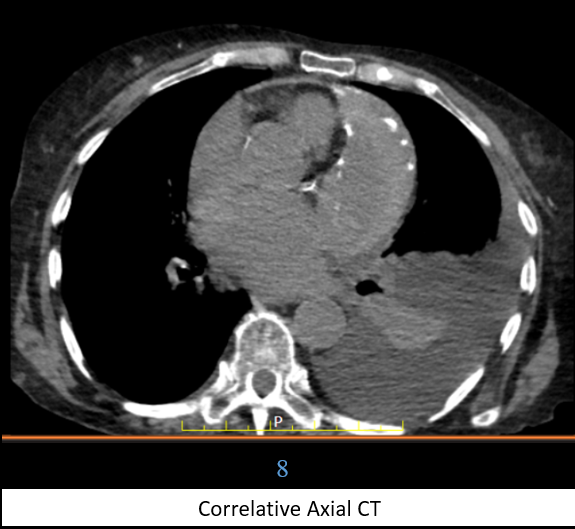
On follow up of patient with PET-CT lesion shows hyperdense mass with peripheral calcification in left pericardium with no uptake abnormal metabolic activity which correlated with previous diagnosis and ruled out any mass/tumor like lesion.
Discussion:
- When any mass is localized to the pericardium, the differential diagnosis includes hematoma, pseudoaneurysm, pericardial cyst, and neoplasm.
- The composition and MRI appearance of pericardial hematomas depends in part on their age.
- It has been reported that acute hematomas are homogeneous, with high signal intensity on both TI-weighted and T2 weighted images, whereas subacute hematomas, between 1 and 4 weeks old, typically demonstrate heterogenous signal intensity, with some areas of high intensity on both TI-weighted and T2-weighted sequences.
- Chronic organized hematomas are of low homogeneous or heterogeneous intensity. They may contain low-intensity foci or have a dark peripheral rim on TI-weighted and gradient echo sequences. These low-intensity areas may reflect calcification.
- Hematomas do not enhance and have no demonstrable internal blood flow on VEC MRI.
- A coronary or ventricular pseudoaneurysm may have an appearance on MRI similar to that of a hematoma. A pseudoaneurysm has a narrow neck with discontinuity in myocardial wall is expected to show internal flow signal on VEC MRI. Detection of blood flow within the mass is important because some paracardiac masses may represent ventricular or coronary artery pseudoaneurysms.
- VEC MRI can identify flow and therefore can differentiate pseudoaneurysm from hematoma.
- Pericardial cysts are well-defined lesions that usually demonstrate homogeneous low signal intensity on T1-weighted images, homogeneous high signal intensity on T2-weighted images, and no enhancement after administration of gadolinium chelates.
- Increased protein concentration in some cysts may result in bright signal intensity on T1-weighted images.
- Neoplasms usually enhance significantly after administration of MR contrast media, although fibromas may show no significant enhancement or characteristic peripheral enhancement with poor or no central enhancement .
- Pericardial hematomas have a variety of etiologies, including trauma, hemorrhagic pericarditis, myocardial or arterial rupture, aortic dissection, and iatrogenic causes.
- Iatrogenic causes of hematoma include anticoagulation, cardiac surgery, and cardiac catheterization.
- Because of its multiplanar capability, contrast resolution, and ability to detect flow, MRI is ideally suited for evaluation of pericardial masses.
Case from literature:

References:
- Watson, W. D., Ferreira, V. M., Sayeed, R., & Rider, O. J. (2019). Serial Cardiac Magnetic Resonance of an Evolving Subacute Pericardial Hematoma. Circulation: Cardiovascular Imaging, 12(12). https://doi.org/10.1161/circimaging.119.009753
- Mustafa Ajam, Manmohan Singh, Abdelrahman Ali, Zachary Elder, Rasikh Ajmal, Ahmed Rashed, Mohamed Shokr. Multimodality Imaging of Pericardial Hematoma. Cardiology and Cardiovascular Medicine 5 (2021): 172-181.
Dr. Srinivas P
Junior Consultant Radiologist
Manipal Hospital Radiology Group (MHRG)
Manipal Hospital Varthur Road
Bengaluru
Dr Deepali Saxena
Senior Consultant Radiologist &
Lead Cardiothoracic Imaging
Manipal Hospital Radiology Group (MHRG)
Manipal Hospital Varthur Road
Bengaluru
