
55 year old female presented with Swelling in medial aspect of the thigh since 10 - 15 days associated with redness.
- 55 year old female presented with Swelling in medial aspect of the thigh since 10 - 15 days associated with redness.
- H/o pedal edema in bilateral legs since 8 years
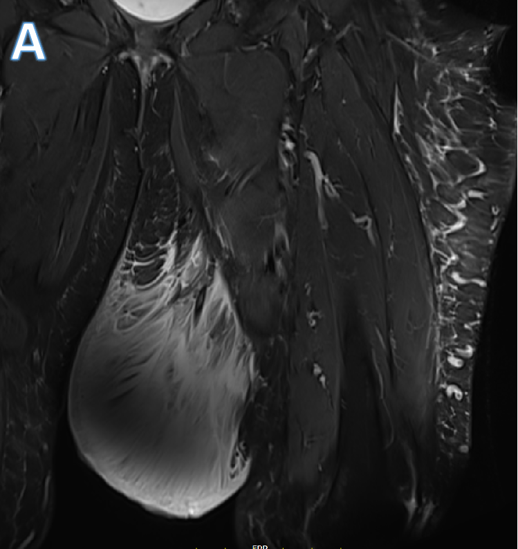
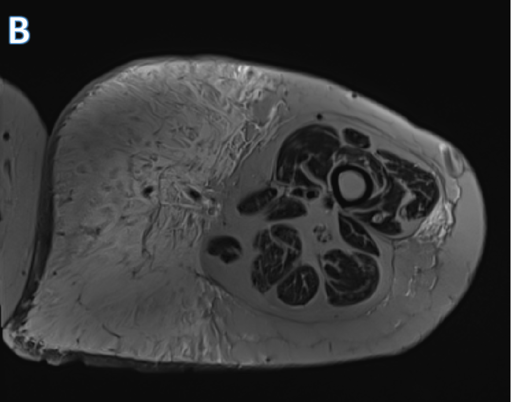
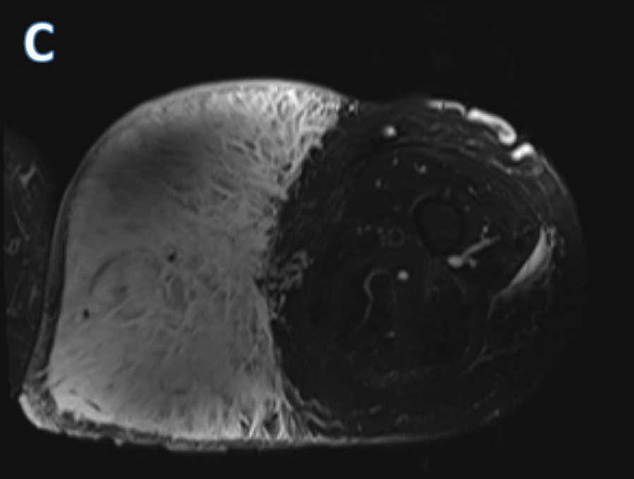
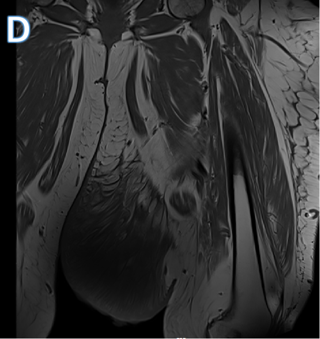
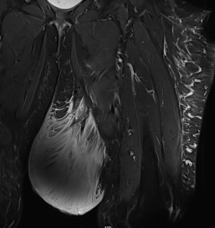
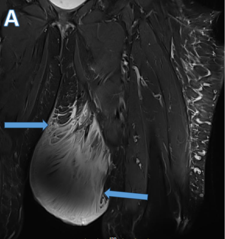
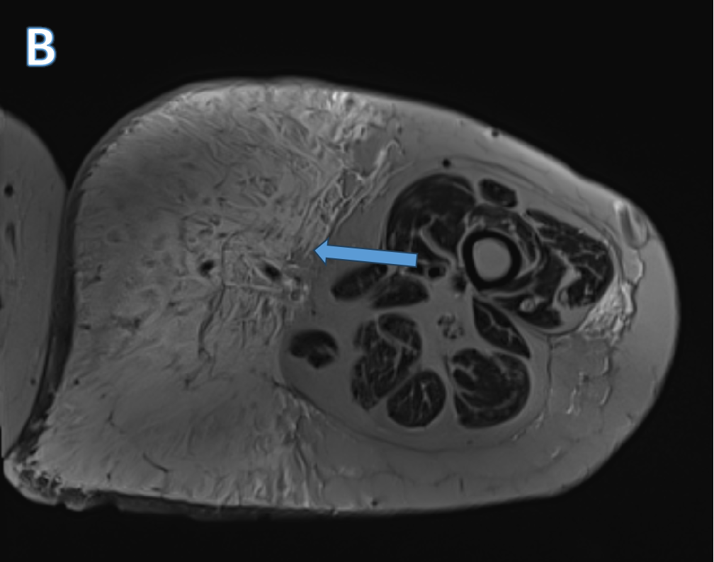
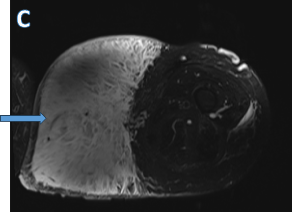
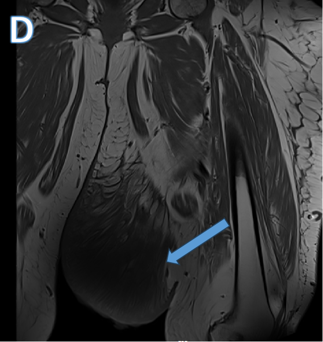
- A pedunculated, adipose matrix in medial thigh which involved the skin and subcutaneous tissue without compromising the fascia or the adjacent muscle planes, appearing T2/STIR hyperintense and T1 hypointense with inflammatory changes. The dermis looked thickened and edematous.
- Axial FS T2 image showing localised edematous changes in the medial aspect of the thigh. Coronal T1 weighted image showing corresponding T1 hypointensity.
DIAGNOSIS
- Massive localised lymphedema ( MLL)
DISCUSSION
Massive localised lymphoedema
- A rare condition , typically present as pendulous masses of adipose tissue ranging in size from 33 to 50 cm in diameter.
- Morbidly obese individuals as a result of localized lymphatic obstruction.
- MLL lesions tend to be slow-growing, enlarging over a period of 1– 10 years. Bilateral masses have been described, but are uncommon
- Most often thigh, other reported sites include the popliteal fossa, scrotum, suprapubic, inguinal region and abdomen.
- Predisposing factors include significant blunt trauma to the inner thigh or chronic lymphoedema resulting from axillary or inguinal lymphadenectomy as well as from vein-stripping, significant weight loss (68–90 kg) following bariatric surgery, inguinal hernia repair and multiple groin operations for idiopathic scrotal oedema, and hemiparesis.
- Infection and skin ulceration have been reported presumably due to mechanical dermal abrasion.
- Clinical examination typically demonstrates a large non-tender swelling with a characteristic peau d’orange appearance of the skin with brawny, non-pitting oedema.
Imaging
- In lymphedema, MR imaging characteristically shows diffuse dermal thickening and oedema within the subcutaneous fat fibrous septae, but unlike in MLL, there is no sharp demarcation between the adjacent subcutaneous tissues at the site of an anatomical distortion, nor is there typical oedema within the fibrous septae fanning out from the pedicle of the lesion.
- Imaging following intravenous administration of gadolinium showed only mild enhancement in the dermis.
Reference
- Khanna, M., Naraghi, A., Salonen, D., Bhumbra, R., Dickson, B. C., Kransdorf, M. J., & White, L. M. (2011). Massive localised lymphoedema: clinical presentation and MR imaging characteristics. Skeletal Radiology, 40(5), 647–652. https://doi.org/10.1007/s00256-010-1080-4
DR DEEPTI H V
Senior Consultant MHRG
DR ANKIT KATARIA
Cross sectional fellow MHRG
