
47-year-old male patient came with history of abdominal pain for 2 days History of trying summersault in the swimming pool, following which the patient developed abdominal pain
- 47-year-old male patient came with history of abdominal pain for 2 days History of trying summersault in the swimming pool, following which the patient developed abdominal pain.
- On examination - diffuse tenderness +
- CT abdomen and pelvis with contrast is requested

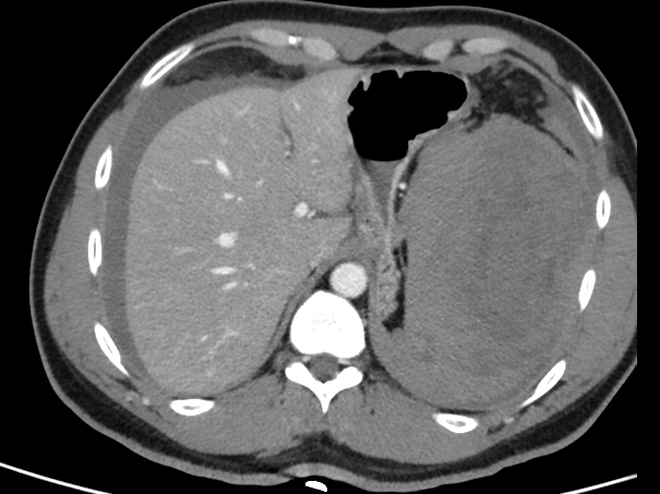

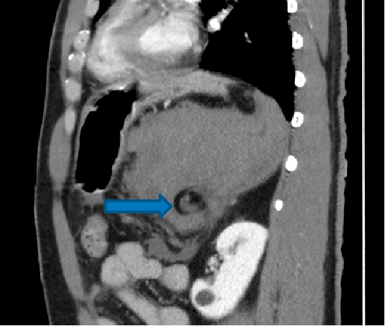
A. Axial arterial and venous phase images shows,
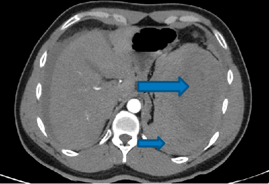
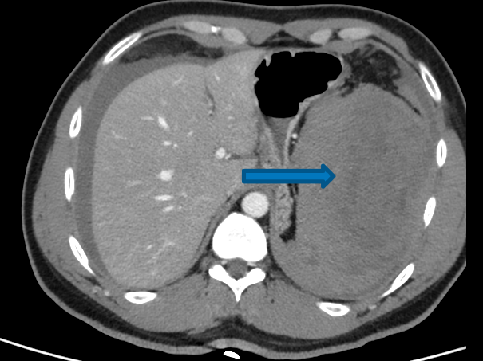
- Spleen is in orthotopic position.
- large subcapsular and perisplenic hematoma.
- Complete non enhancement of splenic parenchyma.
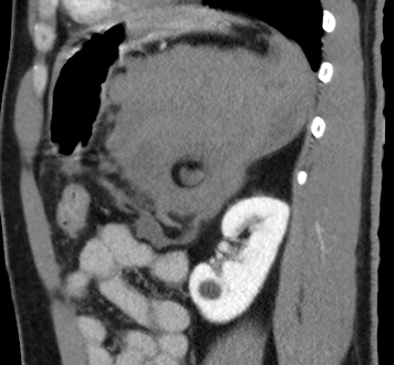
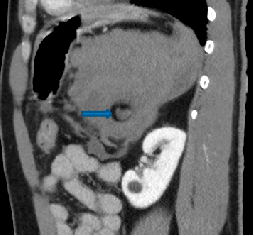
B. Sagittal images shows,
- Twisting of splenic vascular pedicles (whirlpool sign).
DIAGNOSIS
- Splenic torsion causing complete infarction of the orthotopic spleen with splenic rupture.
- Patient underwent laparoscopic splenectomy and peritoneal lavage.
- Intraop findings, Engorged and infarcted spleen was twisted clockwise around its pedicle.
- The spleen was normal in position, located between the stomach and left hemidiaphragm, however, the main splenic ligaments were largely absent.
- Splenectomy was performed.

DISCUSSION
Splenic torsion or volvulus Maybe seen as a complication of wandering spleen due to weakness of splenic ligaments.
Splenic torsion is uncommon in case of orthotopic spleen.
Clinical presentation:
- Abdomen pain / mass / discomfort
Pathology
- Laxity of splenic ligament may lead to wondering spleen
- The spleen can be identified anywhere in the abdomen or pelvis that ultimately result in splenic torsion, congestion, infarction Or in severe cases splenic rupture.
- Ligamentous laxity can be congenital or acquired
- Congenital- failure of the development of normal splenic suspensory ligaments including lieno-renal and gastro-splenic ligaments.
- Acquired- pregnancy (high oestrogen) or trauma.

Ultrasound
- Round hypoechoic solid looking mass in an ectopic or orthotopic location
CT
- Gold standard
- Ectopically located spleen
- Splenic vascular pedicle will demonstrate whirlpool configuration- pathognomonic
- Contrast enhanced CT will demonstrate low density spleen with peripheral enhancement or no enhancement
- Perisplenic free fluid
Treatment
- Open or laparoscopic detorsion and splenopexy is a spleen shaving procedure that can be performed in case of lack of infarction
- In case of splenic infarction- open or laparoscopic splenectomy.
Dr Vikhyath Shetty
Consultant radiologist
Manipal hospital, Yeshwanthpur, Bengaluru.
Dr .Rahul Karthik
Junior Consultant
MHRG,Bengaluru
Dr. Dhinesh kumar T
Radiology resident
Manipal hospital, Yeshwanthpur, Bengaluru.
