
46 year old female, complaints of bony swelling at right calcaneus
46-year-old female, complaints of bony swelling at the right calcaneus
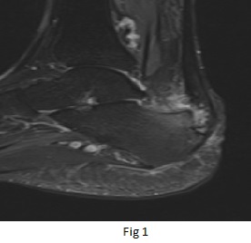
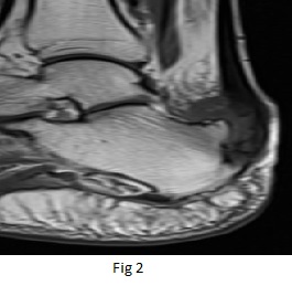
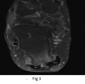
Sagittal PDFS & T1 images (Fig 1 and Fig 2) of ankle joint demonstrate prominence of calcaneus posterosuperiorly with bone edema(green arrow). There is prominent retrocalcaneal bursitis with oedema in the Kager fat pad(white arrow) and insertional tendinopathy of the Achilles tendon(orange arrow). Axial PDFS image(Fig 3) demonstrates partial interstitial tear in the Achilles tendon at distal end(blue arrow). Mild oedema in overlying soft tissues
DIAGNOSIS:
Haglund’s syndrome.
DISCUSSION:
RELEVANT ANATOMY:
- Achilles tendon injuries are common in both athletes and non-athletes and often result from a combination of factors that include overuse, altered biomechanics, and tendon degeneration.
- The Achilles tendon forms from the fusion of the aponeuroses of the medial and lateral heads of the gastrocnemius and the soleus muscles. This musculotendinous unit crosses three joints (knee, tibiotalar, and subtalar) making it more susceptible to injury.
- A 3-6 cm segment proximal to the calcaneal attachment is more susceptible to rupture due to its small cross-section and relative hypo vascularity.
Normal MRI Appearance
- The normal Achilles tendon is of low signal intensity on all imaging sequences.
- The anterior and posterior margins of the Achilles tendon are normally parallel with a concave anterior margin over most of its course.
- The retrocalcaneal bursa is a saddle-shaped structure located posterosuperior to the calcaneus situated between the paratenon and the pre-Achilles fat.
- Subcutaneous fat is normally seen between the Achilles tendon and the skin. Absence of fat in this region may be caused by callus or retro-Achilles bursitis
Haglund syndrome
refers to the triad (Haglund triad) of:
- insertional Achilles tendinopathy
- retrocalcaneal bursitis
- retro-Achilles bursitis
This may be associated with a prominent bursal projection of the posterosuperior calcaneus (Haglund’s deformity)
Insertional Tendinopathy
- MRI depicts distal tendon thickening with ill-defined tendon signal heterogeneity. Partial tears demonstrate hyperintense defects on fluid sensitive sequences. Edema within enthesophytes correlates with more acute symptoms.
Retrocalcaneal Bursitis
- On MRI, the retrocalcaneal bursa is distended with fluid signal and appears hyperintense on fluid-sensitive sequences.
- Enlargement of the bursal anteroposterior dimensions to greater than 2mm is consistent with bursitis. There is often an associated element of inflammation in the adjacent paratenon and Achilles tendon insertion. Associated partial Achilles tendon tear, peritendinitis, tendinopathy, or ossification is common.
Retro-Achilles Bursitis
- The retro-Achilles bursa is an adventitious bursa that develops as a response to friction between the posterior aspect of the calcaneus or the Achilles tendon and the skin.
- It is most often seen at the posterolateral aspect of the calcaneus and is frequently associated with tight or poorly fitted shoes with a rigid heel contour.
Dr Naveen SS, MD
Cross-sectional imaging fellow MHRG
Dr Dayanand Sagar G, MD
Consultant Radiologist MHRG
