
45 year old lady, with no known comorbidities presented with periumbilical swelling and generalized abdominal pain for 2 months
- 45 year old lady, with no known comorbidities presented with periumbilical swelling and generalized abdominal pain for 2 months. No prior surgeries.
- On examination – Per abdomen: soft, partially reducible umbilical hernia
- Planned for mesh repair of ventral hernia and underwent pre-op CECT.

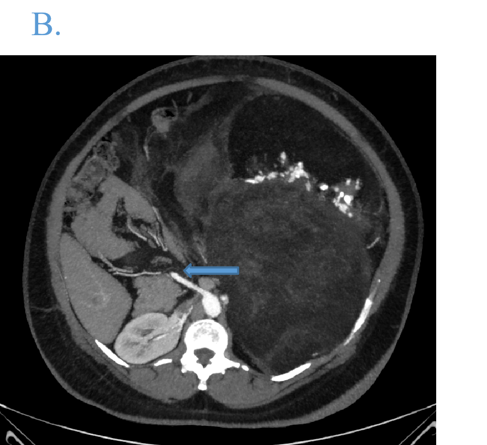
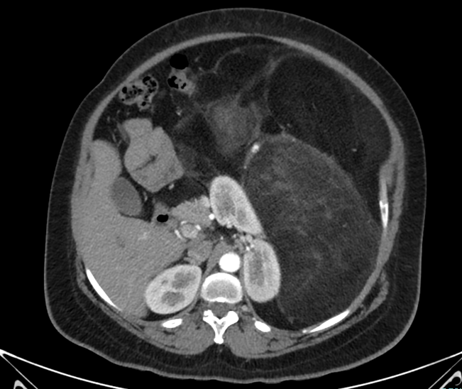
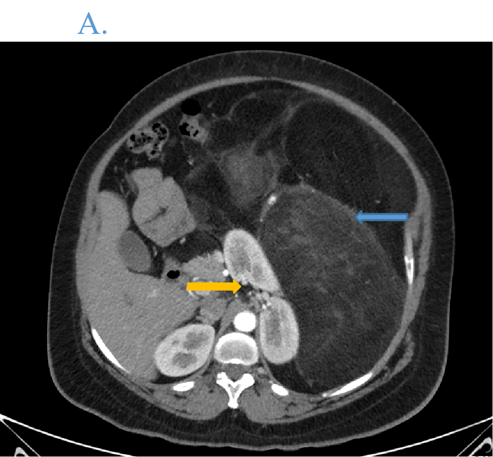
- Axial arterial phase image depicting large retroperitoneal lesion with multiple enhancing septations (blue arrow) in left side of abdomen displacing adjacent organs (yellow arrow).
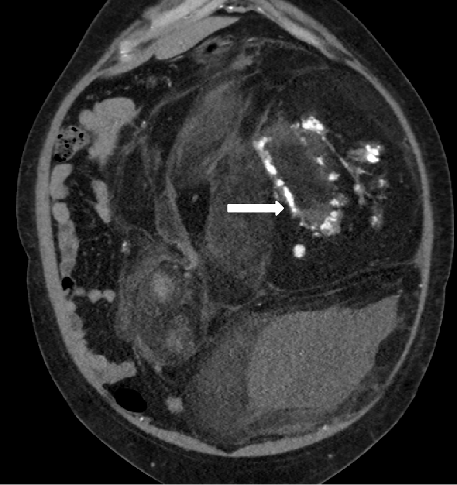
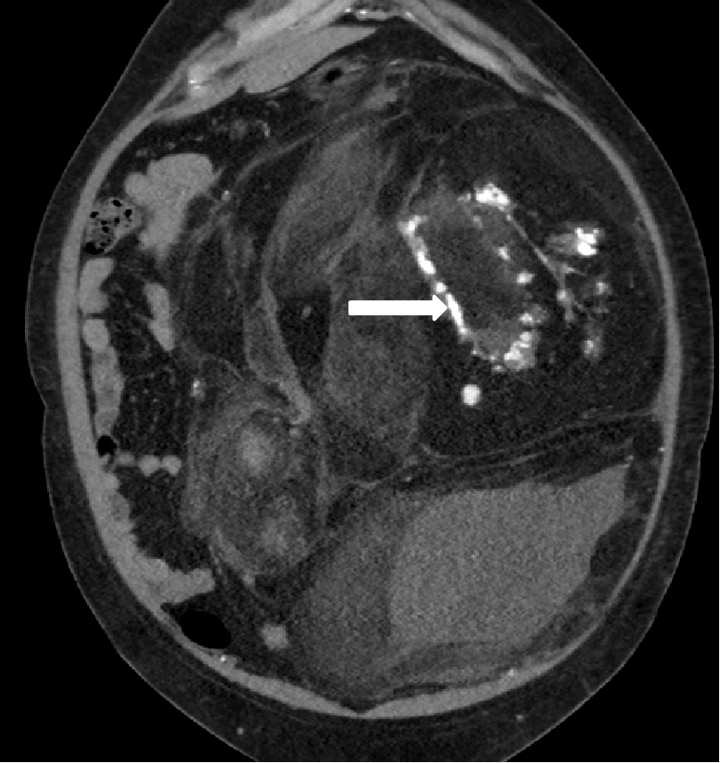
- Coronal image shows that the lesion occupies the abdomen near completely with predominant fat density. Solid enhancing component with calcific specks are seen (white arrow).
- Coronal image shows that the lesion occupies the abdomen near completely with predominant fat density. Solid enhancing component with calcific specks are seen (white arrow).
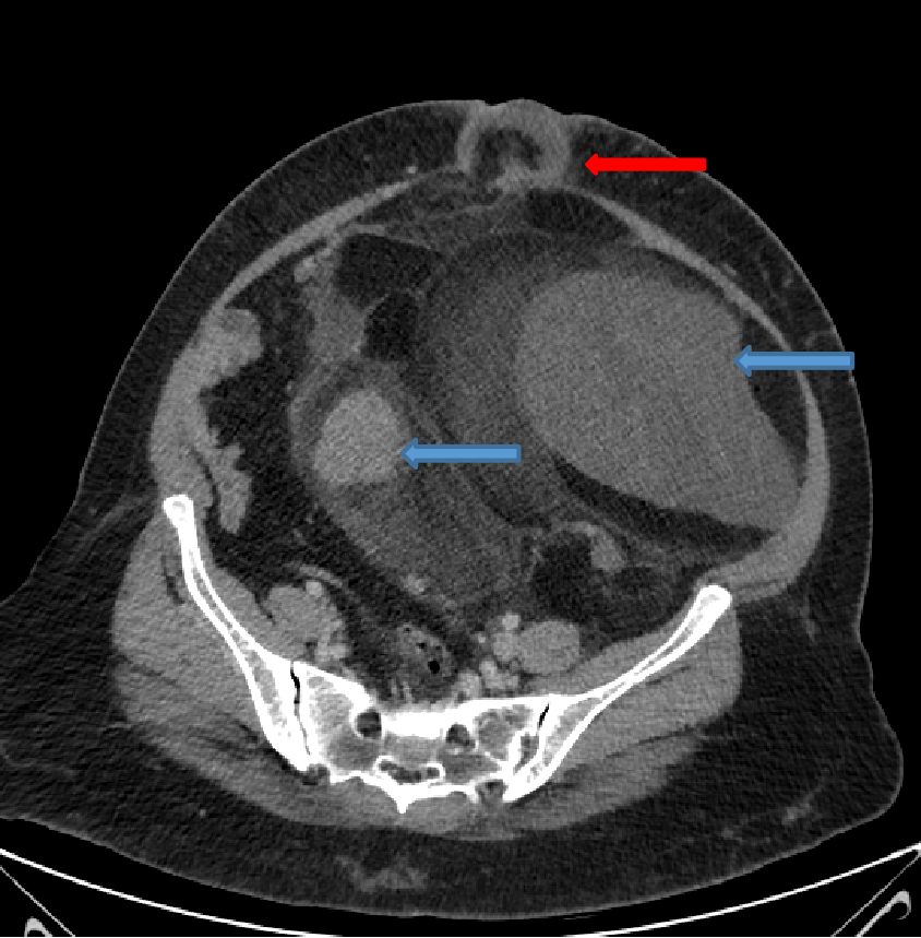
- Axial venous phase image at caudal level shows solid enhancing components within the lesion (blue arrows), and a ventral hernia with fat and fluid within (red arrow).
- Patient underwent resection of the lesion followed by left hemicolectomy. Samples were sent for HPE.


RETROPERITONEAL LIPOSARCOMA (HPE:- Dedifferentiated variant)
DISCUSSION:
- Primary retroperitoneal sarcomas (RPS) represent around 10–16% of all sarcomas, with liposarcomas and leiomyosarcomas being the most common subtypes.
- RPS primarily present as large masses, progressively encasing adjacent structures, causing mass effect.
- Symptoms are commonly associated with displacement, compressive or obstructive phenomena, and include abdominal or back pain, urinary tract obstruction, edema of the lower limbs, nerve compression, and vague digestive symptoms.
- Recurrence - 45 to 49% within the first five years , 60 to 82% at 10 years after initial treatment. Lifelong surveillance is advised.
- Nodal mets is rare and is a poor prognostic sign.
- Retroperitonel liposarcomas have 5 histological subtypes:
- Well differentiated
- Myxoid (myxoid has moer mucopolysacharides – T2 hyperintense)
- Pleomorphic
- Round cell
- Dedifferentiated
- Often metastasize hematogenously.
- Calcifications are often present in dedifferentiated variant and implies poor prognosis.
- Role of radiologist is to rule out other conditions like adenocarcinoma, retroperitoneal fibrosis, lymphoma, paraganglioma etc (LPS mimics angiomyolipoma in having fat density).
- CECT helps to identify the origin, contents (fat, solid, calicific) and also mass effect over adjacent organs with complications if any (eg:- ureteric obstruction or vascular compromise).
- Within the lesion there could be thin septations (low grade) to heterogenous sold mass (high grade).
- MRI – myxoid components can show T2 hyperintensity. Delayed post contrast enhancement can be seen.
TREATMENT
- Surgery is mainstay.
- Resection margins must be clear, otherwise resulting in high rates of recurrence.
REFERENCES
- Mota MM, Bezerra RO, Garcia MR. Practical approach to primary retroperitoneal masses in adults. Radiologia brasileira. 2018 Nov 23;51:391-400.
- Porrello G, Cannella R, Randazzo A, Badalamenti G, Brancatelli G, Vernuccio F. CT and MR Imaging of Retroperitoneal Sarcomas: A Practical Guide for the Radiologist. Cancers (Basel). 2023 May 30;15(11):2985. doi: 10.3390/cancers15112985. PMID: 37296946; PMCID: PMC10252049.
- Craig WD, Fanburg-Smith JC, Henry LR, Guerrero R, Barton JH. Fat-containing lesions of the retroperitoneum: radiologic-pathologic correlation. Radiographics. 2009 Jan;29(1):261-90.
Dr THIMMANNA K.D
Consultant Radiologist
Manipal Hospital, Yeshwanthpur, Bengaluru.
Dr ANAGH VISHNU NARAYANAN
Radiology resident
Manipal Hospital, Yeshwanthpur, Bengaluru.
