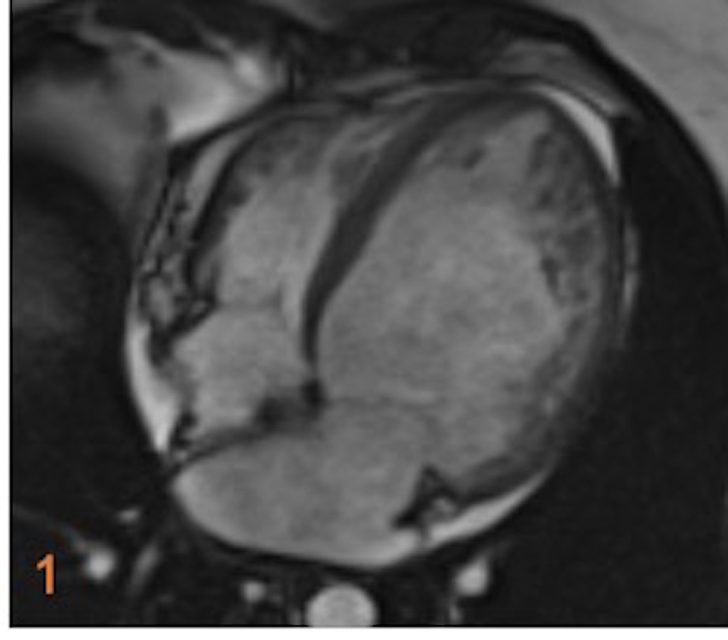
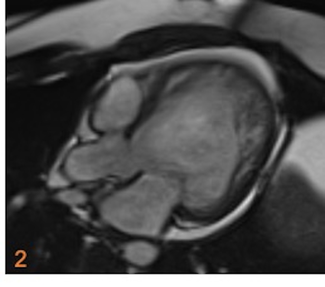
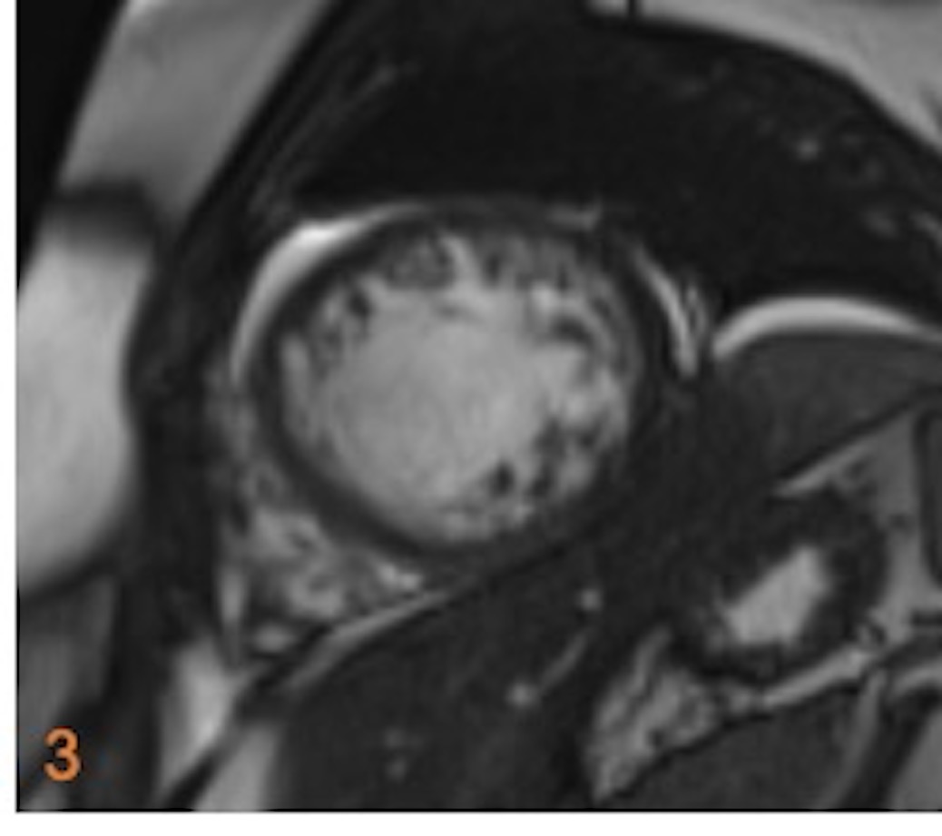
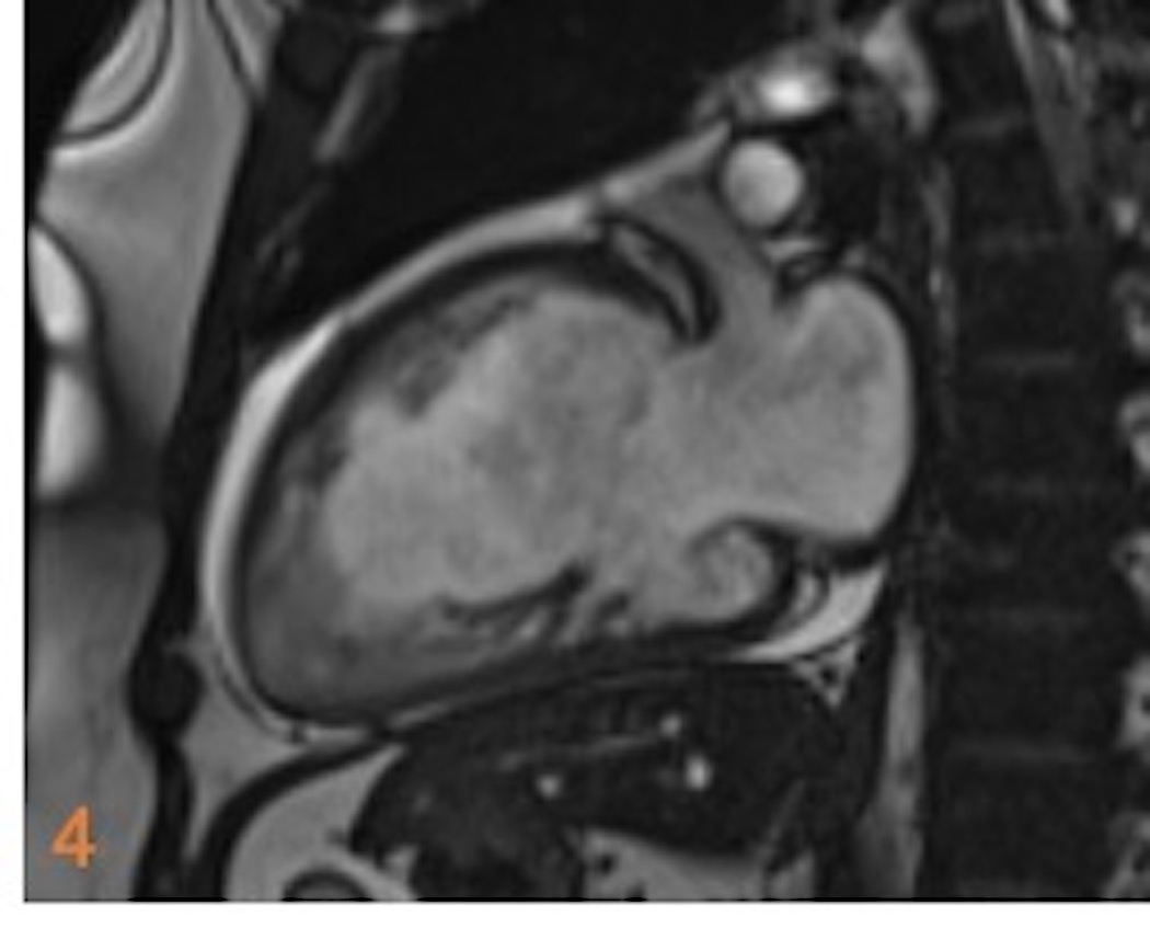
45 year old gentleman presented with worsening heart failure. No prior investigations were done.
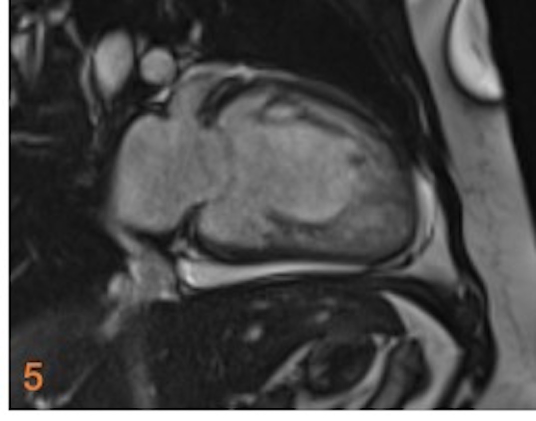
Fig. 1-5. Radial cine SSFP images. Increased trabeculations with increased depth of intertrabecular recesses are seen in Left ventricle (LV). LV Non-compacted to compacted myocardial ratio is 5.7. There is involvement of right ventricle as well in this case.
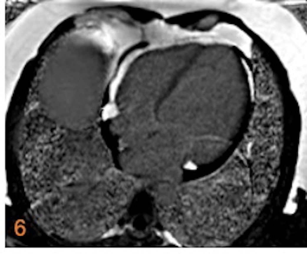
Fig. 6. 4CH PSIR. No myocardial delayed gadolinium enhancement.
DIAGNOSIS:
Non-compaction cardiomyopathy.
DISCUSSION:
- Congenital cardiomyopathy with arrest of compaction in endocardial myocardium in fetal life. The outer layer consists of normally compacted myocardium.
- However, it can be diagnosed at a later age in life when patients get evaluated for etiology for heart failure.
- CMR criteria for LV non-compaction:
Non-compacted to compacted myocardial ratio >2.3 (in end-diastole) Trabecular LV mass >20% of total LV mass and NC indexed mass > 15 g/m2
- Other features: Deep intertrabecular recesses
- Evaluate to rule out:
- Thrombus in LV hidden in deep recesses.
- RV involvement with multiple deep trabeculae.
REFERENCES:
- Dreisbach, J.G., Mathur, S., Houbois, C.P. et al. Cardiovascular magnetic resonance based diagnosis of left ventricular non-compaction cardiomyopathy: impact of cine bSSFP strain analysis. J Cardiovasc Magn Reson 22, 9 (2020). https://doi.org/10.1186/s12968-020-0599-3
- https://www.escardio.org/Journals/E-Journal-of-Cardiology-Practice/Volume-10/Left-ventricularnoncompaction#:~:text=Criteria%20for%20diagnosis%20by%20CMR,and%20specificity%20of%2099%25).
Dr. Deepali Saxena.
DNB, Fellowship Cardiothoracic Imaging (USA
Lead Cardiac Imaging
Manipal Hospitals Radiology Group.

