42 years old female with chronic headache, difficulty in chewing, swallowing and dysarthria
42 years old female with chronic headaches, difficulty in chewing, and swallowing, and dysarthria
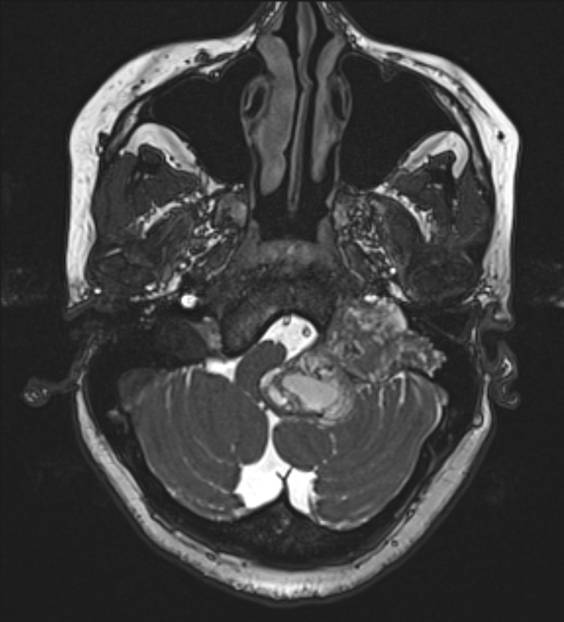

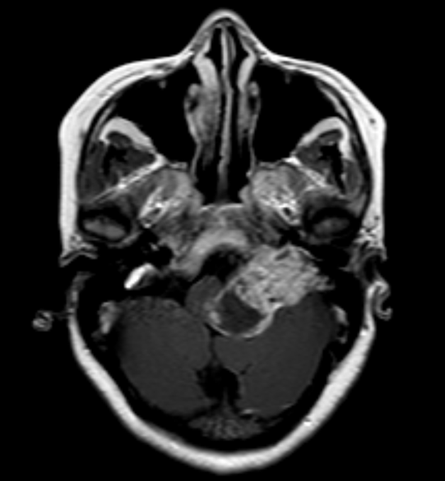
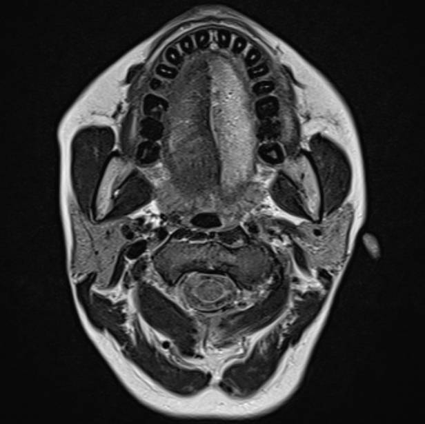
There is a lobulated extra-axial heterogenous signal mass lesion centered in left hypoglossal canal (orange arrow) causing remodelling of left petrous bone. Right hypoglossal canal shown for comparison (blue arrow Fig1). Multiple foci of susceptibility are seen within the lesion (Fig 2). There is heterogenous enhancement (Fig 3) with few non enhancing cystic- necrotic areas. Features of left hypoglossal palsy with fatty changes and volume loss seen in left hemitongue (Fig 4).
Diagnosis:
HYPOGLOSSAL SCHWANOMMA
Discussion:
Hypoglossal schwannomas are uncommon and presents with ipsilateral hypoglossal nerve palsy
Clinical presentation: dysarthria, hemitongue atrophy, tongue fasciculation & deviation towards the side affected.
Key Diagnostic Features: Heterogeneously enhancing mass with sometimes dumbbell shaped seen centered in hypoglossal canal causing its smooth widening. Associated features of hypoglossal denervation changes with ipsilated tongue atrophy and fatty replacement
Differential diagnosis:
-
- Meningioma: Dural tail, hyperostosis, calcification, and intense homogeneous enhancement
- Schwannoma from other cranial nerves (vestibular, glossopharyngeal): The diagnosis will depend on which cranial foramen or canal the lesion is centered and widened
- Glomus jugulare or jugulotympanicum: Permeative pattern of bone destruction instead of bone remodeling, salt and pepper pattern with multiple hypertrophied feeding vessels
- Metastasis: Bone destruction, multiplicity, and relevant clinical history
Rx: Resection / stereotactic radiosurgery.
Dr. Sriram Patwari
MD, PDCC (Neuroradiology)
Consultant Radiology, Co-lead Neuroradiology
Manipal Hospitals Radiology Group
Dr. Sushant Mittal
MD
Cross-sectional fellow,
Manipal Hospitals Radiology Group