
41 year old male patient with history of sharp shooting pain in left sole radiating to 1st and 2nd toe. 25yr old history of local regional trauma (via glass)
- 41 year old male patient with history of sharp shooting pain in left sole radiating to 1st and 2nd toe. 25yr old history of local regional trauma (via glass).
- Tenderness present at plantar aspect of left foot.
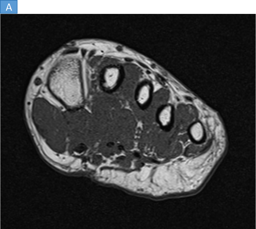
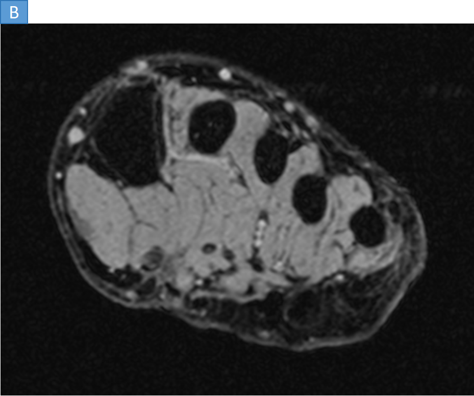
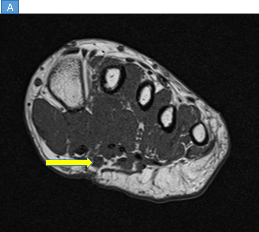
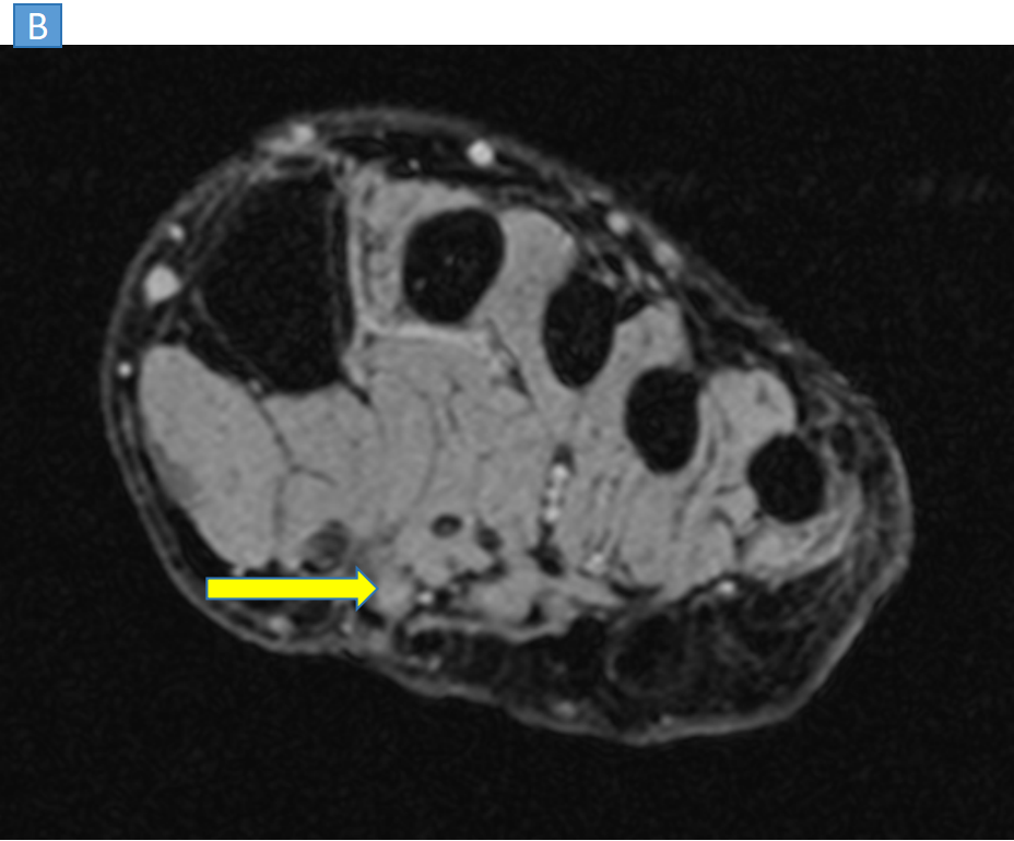
- AXIAL T1 (IMAGE- A) and PD FS (IMAGE- B) image shows well defined small lesion T1 hypointense and PD FS hyperintense lesion along the plantar aspect in between the proximal thirds of 1st and 2nd metatarsals . Cutaneous irregularity is also noted along the planter aspect at the site of prior trauma.
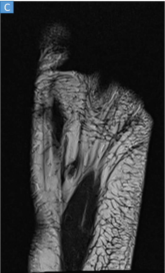
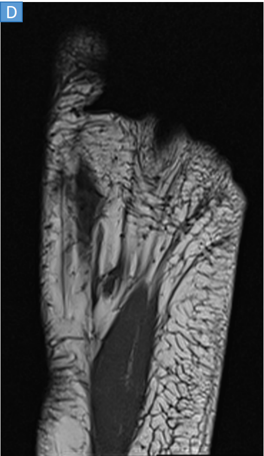
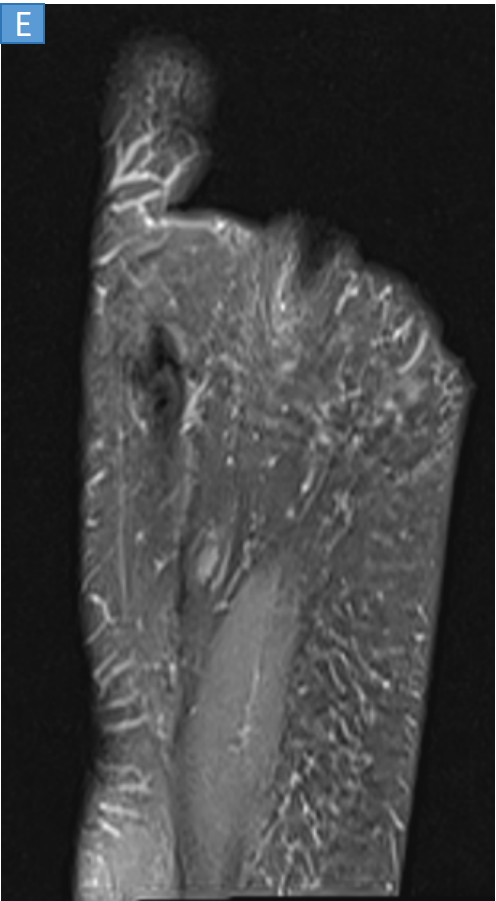
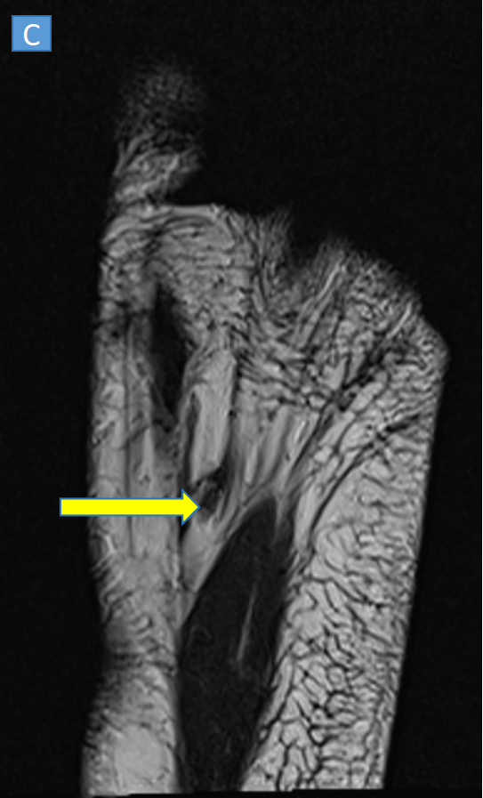
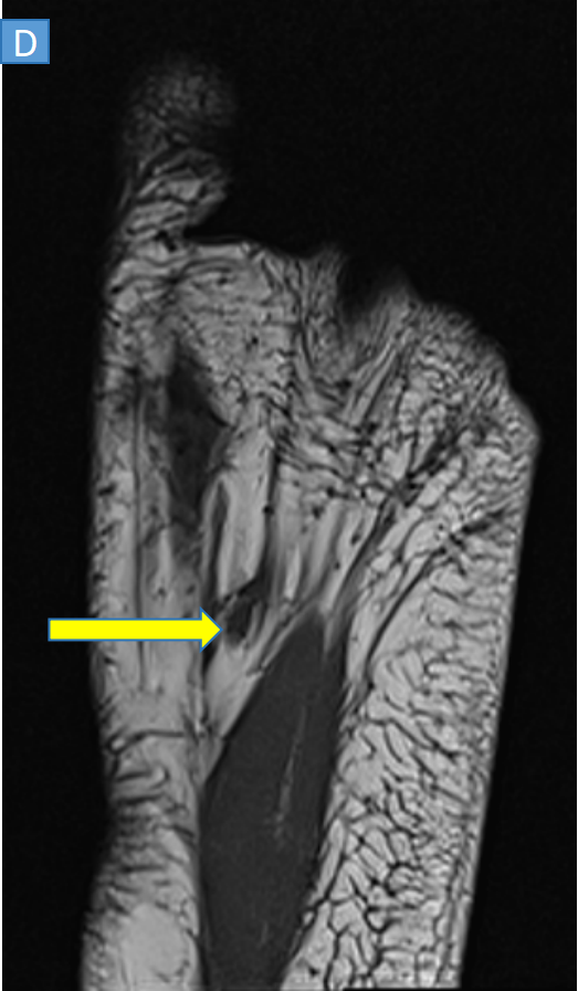
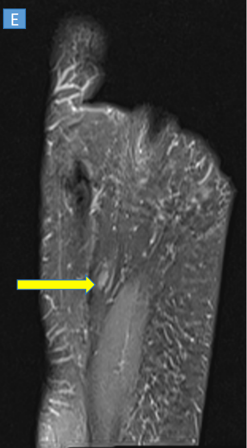
- CORONAL T2 (Image C), T1 (Image D), PD FS (Image F) shows well defined small lesion T2 isointense,T1 hypointense and PD FS hyperintense lesion along the plantar aspect in between the proximal thirds of 1st and 2nd metatarsals .
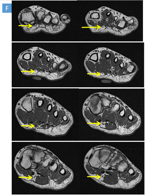
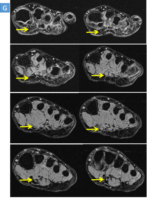
- AXIAL T1 (IMAGE F) and and PD FS (IMAGE G)- shows the lesion involving the plantar nerve.
DIAGNOSIS
- Traumatic neuroma along branch of medial plantar nerve in the 1st webspace.
DISCUSSION
- Traumatic neuroma is a non-neoplastic lesion that occurs in response to injury or previous surgery. It is a hyperplastic, reparative nerve reaction after injury and typically manifests as a nodular mass.
- On MRI It is hypointense to the muscle on T1, isointense on T2 and hyperintense on PDFS.
- Course of plantar nerve in foot.
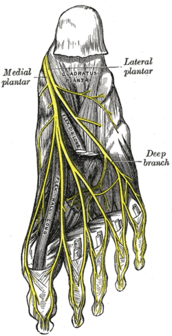
- Median plantar nerve (branch of the Tibial nerve) at its origin, under the laciniate ligament passes deep to the abductor hallucis muscle and re-appearing between this muscle and the flexor digitorum brevis.
- The lateral plantar nerve (branch of the Tibial nerve) travels obliquely to the lateral side of the foot deep to the flexor digitorum brevis.
Differential Diagnosis:
MORTON'S NEUROMA:
- Thickening and degeneration of one of the interdigital nerves of the foot.
- Most commonly between the 3rd and 4th metatarsal heads.
- More common in woman.
- C/O Sharp shooting pain that is worse on standing characteristically begins in the sole of the foot and radiates to the exterior.
NEUROFIBROMA:
- 20 and 30 years of age.
- Superficial painless lesion seen in the soft tissue, sometimes without evidence of an origin from a peripheral nerve.
BENIGN SCHWANNOMA:
- Encapsulated nerve sheath tumor that consists histologically of two components cellular and myxoid.
- Predilection for the head, neck, and flexor surfaces of the upper and lower extremities.
REFERENCES
- Andrew L. Folpe, in Diagnostic Surgical Pathology of the Head and Neck (Second Edition), 2009
- https://radsource.us/traumatic-neuroma/
- Orthopaedic Pathology, 5th Edition, by Peter G. Bullough, MB, ChB.
Dr. Ritika Chamadia
Consultant Radiologist,
Manipal Hospital, Kharadi, Pune.
Dr. Madhuri and Dr. Aditya
Radiology Residents,
Manipal Hospital,Kharadi, Pune.

