
4-month-old child with developmental delay and abnormal eye movements.
4-month-old child with developmental delay and abnormal eye movements.
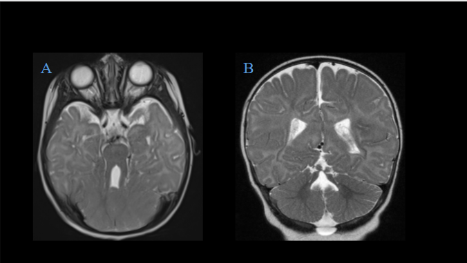
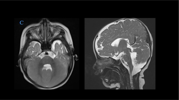
- Representative T2W axial images demonstrates thickened bilateral superior cerebellar peduncles giving the molar tooth appearance.
- Representative T2W coronal images demonstrates hypoplastic and dysplastic vermis with abnormal sulcation in cerebellar hemispheres.
- Representative T2W axial and coronal images demonstrates deformed roof and fastigium of fourth ventricle and having bat wing shaped configuration on axial images.
- Literature image of color-coded FA DTI maps at the level of the superior cerebellar peduncles demonstrates:
- Normal vertical orientation of fibers in healthy subjects (Images on the left) within the superior cerebellar peduncles, characterized by a blue color coding and decussation of the superior cerebellar peduncles (red dot) at the level of the inferior colliculi of the midbrain.
- In Joubert syndrome (Images on the right) there is a horizontal orientation of fibers in the superior cerebellar peduncles, characterized by the green color coding and absence of the “red dot” within the midbrain confirms the failure of the superior cerebellar peduncles to decussate.
DIAGNOSIS:
JOUBERT SYNDROME AND RELATED DISORDERS
DISCUSSION:
- Joubert Syndrome is a rare autosomal recessive condition characterized by agenesis of the cerebellar vermis, and patients typically present with episodic hyperpnea, irregular eye movements, ataxia, and intellectual disability.
- The imaging hallmark of JS is the molar tooth sign. This appearance of the midbrain is attributed to the combination of dysplasia of the cerebellar vermis, foreshortened midbrain, and thickened and elongated superior cerebellar peduncles.
- Molar tooth sign is not unique to Joubert syndrome and has been reported in a number of other conditions. The term Joubert Syndrome and Related Disorders (JSRD) is used to describe this heterogeneous group of disorders that show the molar tooth sign on radiologic imaging.
- A midline cleft between the cerebellar hemispheres is seen resulting in a batwing appearance of the fourth ventricle on axial images.
- Absence of fiber decussation in the superior cerebellar peduncles and pyramidal tracts , which can be assessed by diffusion tensor imaging.
- Abnormal inferior olivary nucleus.
References:
- Poretti A, Boltshauser E, Loenneker T, Valente EM, Brancati F, Il’Yasov K, Huisman TA. Diffusion tensor imaging in Joubert syndrome. American journal of neuroradiology. 2007 Nov 1;28(10):1929-33.
- Poretti A, Huisman TA, Scheer I, Boltshauser E. Joubert syndrome and related disorders: spectrum of neuroimaging findings in 75 patients. American journal of neuroradiology. 2011 Sep 1;32(8):1459-63.
Dr. Sriram Patwari
MD, PDCC (Neuroradiology), EDiNR
Consultant Radiologist
Manipal Hospital, Yeshwanthpur, Bengaluru
Dr Akshay K
Radiology resident
Manipal Hospital, Yeshwanthpur, Bengaluru
