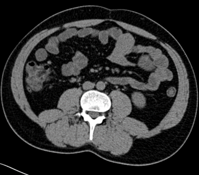
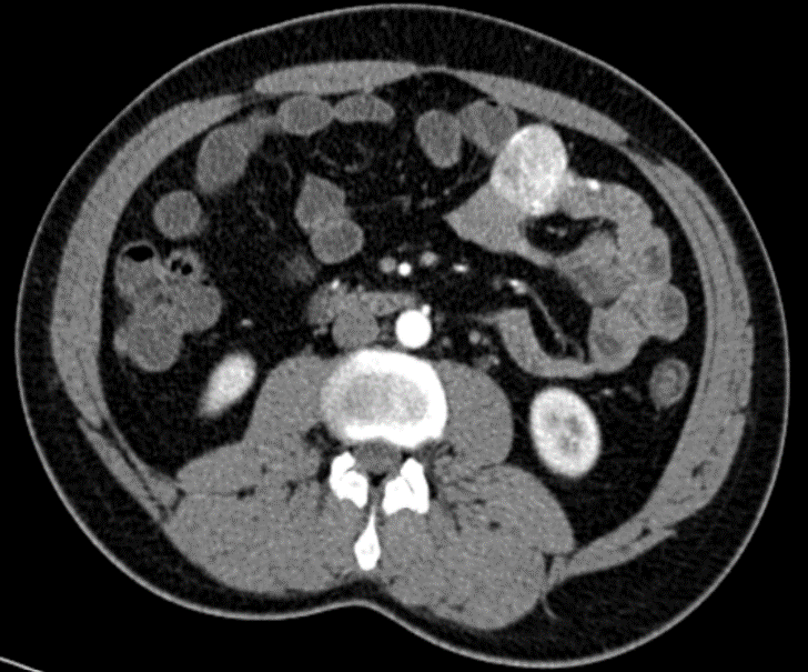
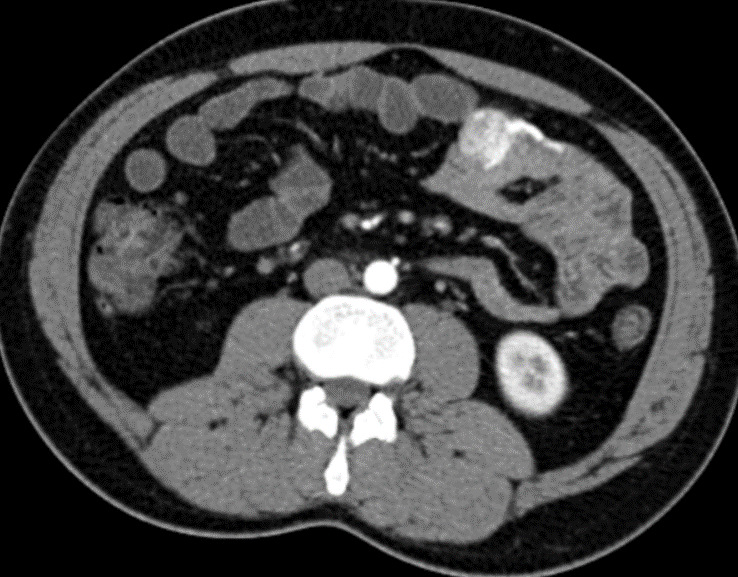
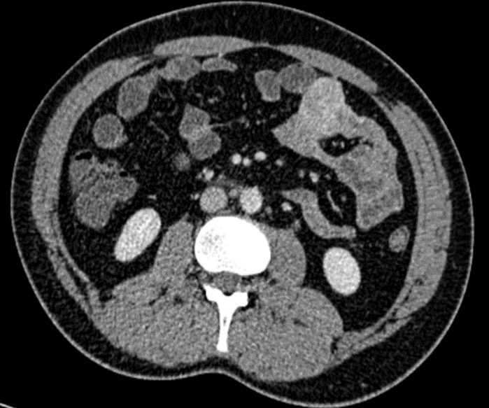
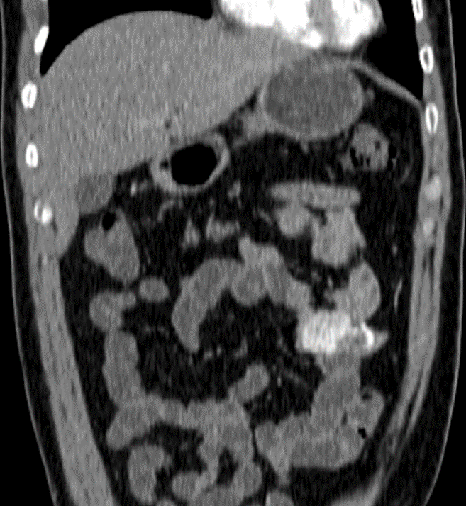
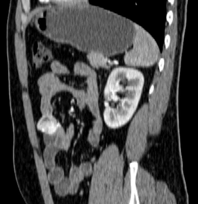
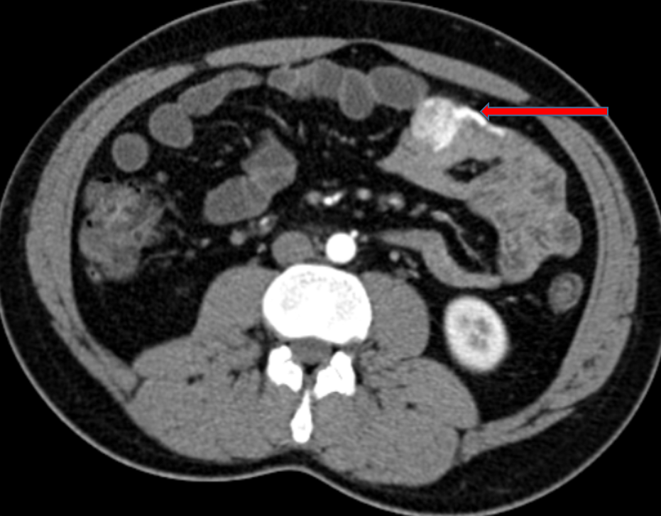
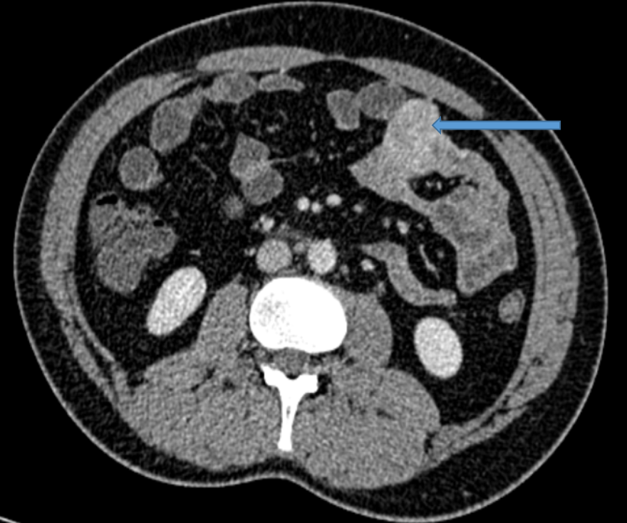
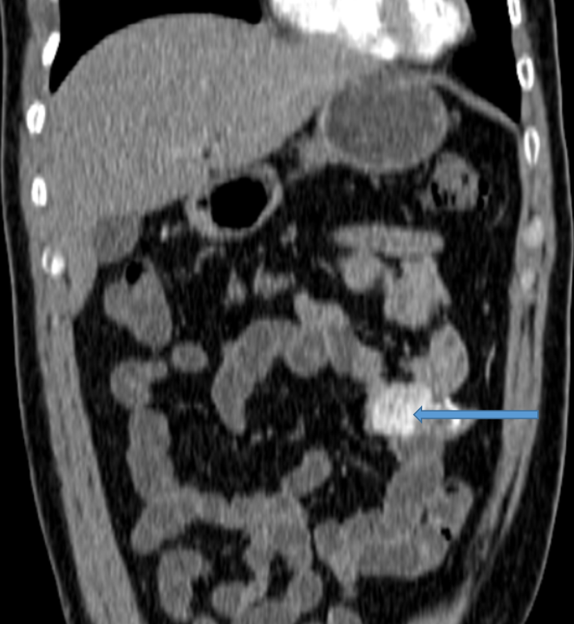
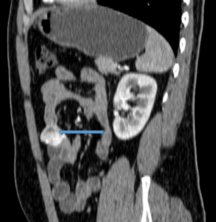
37y Male, presenting with PR bleeding which is on/off since 2 years.
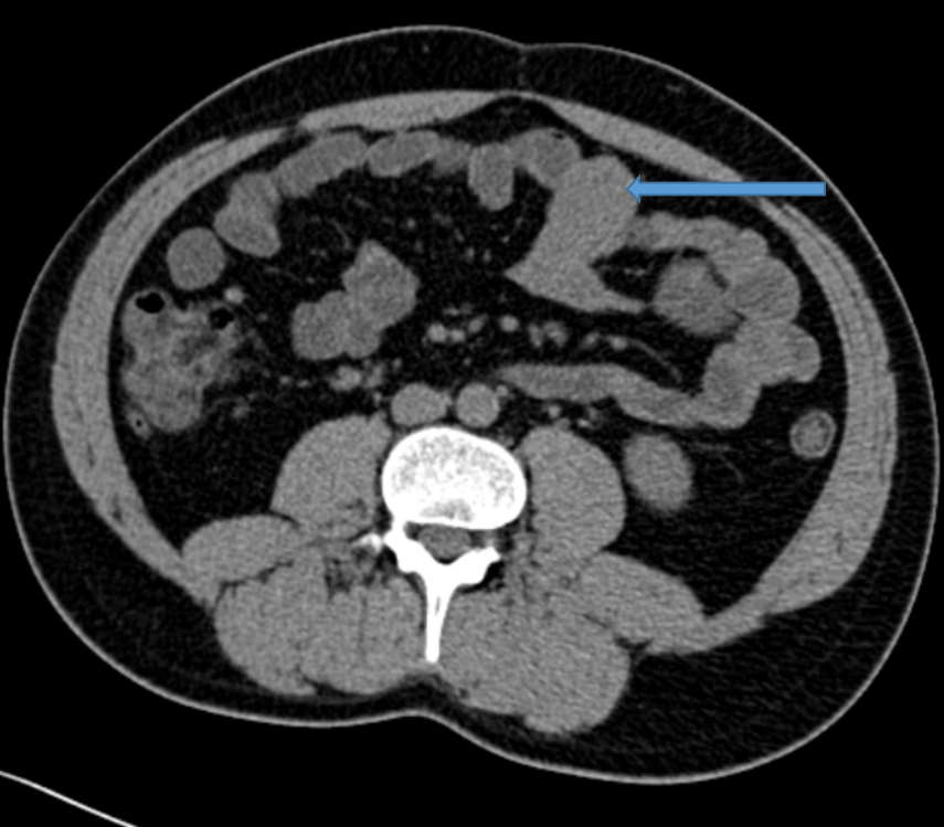
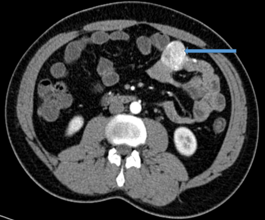
Blue Arrow:
- Relatively well-defined soft tissue predominantly exophytic lesion in the mid left abdominal small bowel involving the jejunal loop.
- It demonstrates intense enhancement in the arterial phase and relative washout in the venous phase.
- No evidence of calcifications.
- No adjacent desmoplastic reaction.
Red arrow:
- The lesion is supplied by jejunal branches of the superior mesenteric artery, which appear hypertrophied.
No evidence of any bowel wall thickening or bowel obstruction
DISCUSSION:
- Also known as small bowel neuroendocrine tumors (SBNETs)
- The most common gastrointestinal neuroendocrine tumors.
- Most frequently involve the terminal ileum.
Epidemiology
- Small bowel carcinoids account for ~40% of gastrointestinal neuroendocrine tumors.
Markers
- 5-HIAA (5-hydroxyindoleacetic acid): usually suggests a functioning carcinoid tumor.
- Chromogranin A (CgA): considered a valuable tool in the diagnosis of neuroendocrine neoplasia in genera.
Clinical presentation
- Small bowel carcinoids are slow growing and present with vague symptoms:
- Weight loss
- Fatigue.
- Diarrhoea.
- Abdominal pain
More specific symptoms include:
- carcinoid syndrome
- small bowel obstruction.
- Haematochezia.
- obstructive jaundice: in duodenal carcinoid
Location
Gastrointestinal tract carcinoid(60-85% of all carcinoids)
- Small bowel: ~40% of gastrointestinal carcinoids, mostly in the terminal ileum
- Rectum (~22.5%)
- Colon (~15%)
- Appendix(~10%)
- Stomach (~7.5%)
- Pancreas (~7.5%)
Carcinoid tumors of the lung(~25% of all carcinoids)
- Bronchial carcinoid
- Peripheral pulmonary carcinoid tumors
Primary hepatic carcinoid
Ovarian carcinoid: accounts for 0.5% of carcinoid tumours and 0.3% of ovarian tumours.
Thymic carcinoid.
Pathology
- Carcinoid tumors are neuroendocrine tumors arising from APUD cells.
- They can cause a desmoplastic reaction in nearby tissue, leading to fibrosis and tethering of the adjacent bowel.
- The primary tumor in small bowel carcinoid is typically only up to 3.5 cm in size.
- Metastases, commonly to the mesentery, liver, and lymph nodes, often exceed the size of the primary neoplasm.
- Multiple primaries and metachronous tumors in other organs can often occur.
- About 45% (range 30-60%) of patients with small bowel carcinoid have metastatic disease at presentation.
- The larger the primary, the greater the likelihood of metastases.
Radiographic features
CT
- Primary small bowel carcinoids are not always seen on imaging given their small size.
- Targeted protocols including a late arterial phase and CT enterorrhaphy improve the sensitivity for detecting small bowel tumors.
Characteristics of the primary lesions on CT include:
- Polypoid or plaque-like appearance.
- Hyper enhancing on arterial phase.
- Can cause distortion and focal fixation of the affected small bowel loop – “hairpin” kinks in the course of the small bowel.
- Calcifications are present in up to 70% of cases.
Mesenteric metastases can appear well-defined or spiculated on CT, with stranding due to fibrosis and desmoplastic reaction (leading to a characteristic “spoke-like” appearance of mesenteric vessels).
In the liver, metastases strongly enhance in the arterial phase due to their vascularity, then become in the setting of dense to liver parenchyma in the delayed phase.
Nuclear medicine
- Gallium-68 octreotide PET-CT (e.g. Ga-68 DOTATATE) has shown improved accuracy for detection of neuroendocrine tumors relative to indium-111 pentetreotide (Octreoscan) SPECT/CT.
- Indium-111 octreotide (e.g. Octreoscan) SPECT-CT.
- Iodine-123 MIBGwill also concentrate on carcinoid tumours, including the low percentage (~15%) that are negative with indium-111 octreotide.
Treatment and prognosis
- Surgical resection is considered the main treatment for localized tumors. Carcinoid syndrome mainly treated by somatostatin analog therapy.
- Everolimus is used as 2ndline treatment.
- Surgical resection, embolization and ablation can be used to treat hepatic metastases.
REFERENCES
- Levy AD, Sobin LH. From the archives of the AFIP: Gastrointestinal carcinoids: imaging features with clinicopathologic comparison. Radio graphics : a review publication of the Radiological Society of North America, Inc. 27 (1): 237-57. doi:10.1148/rg.271065169 – Pubmed
- Strosberg JR, Nasir A, Hodul P et-al. Biology and treatment of metastatic gastrointestinal neuroendocrine tumors. Gastrointestinal cancer research : GCR. 2 (3): 113-25. Pubmed
- Ganeshan D, Bhosale P, Yang T, Kundra V. Imaging Features of Carcinoid Tumors of the Gastrointestinal Tract. AJR Am J Roentgenol. 2013;201(4):773-86. doi:10.2214/ajr.12.9758 – Pubmed
- Horton KM, Kamel I, Hofmann L et-al. Carcinoid tumours of the small bowel: a multi-technique imaging approach. AJR. American journal of roentgenology. 182 (3): 559-67. doi:10.2214/ajr.182.3.1820559 – Pubmed
- Larouche V, Akirov A, Alshehri S et-al. Management of Small Bowel Neuroendocrine Tumours. (2019) Cancers. doi:10.3390/cancers11091395 – Pubmed.
Dr. Vishwanath Joshi
Consultant Radiologist.
Manipal Hospital Radiology Group (MHRG)
Manipal Hospital, Bengaluru.
Dr. Srinivas P
Fellow in Radiology
Manipal Hospital Radiology Group (MHRG)
Manipal Hospital, Bengaluru.

