36 year old gentleman with history of left hip pain since 3 weeks
- 36 year old gentleman with history of left hip pain since 3 weeks.
- On examination reduced ROM at hip joint and tenderness over gluteal area.
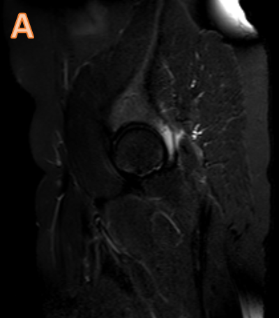
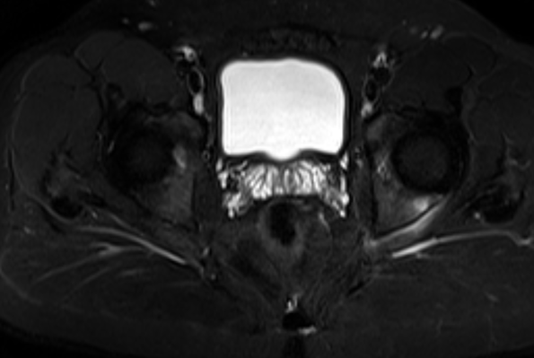
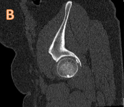
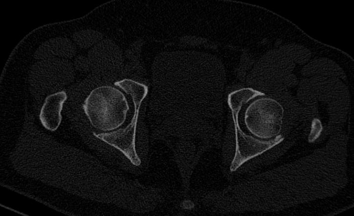
IMAGE A: Sagittal and axial STIR images show marrow edema at posterior wall of left acetabulum with traces of periosteal edema/fluid along posterior margin of acetabulum.
IMAGE B: CT demonstrates small bone fragment abutting the posterior lip of left acetabulum in hip joint space with a tiny adjacent lytic area.
DIAGNOSIS:
Inta-articular osteoid osteoma
Discussion:
- Osteoid osteomas are benign bone forming tumors that tend to occur in patients between the ages of 10 and 30 years.?
- Osteoid osteomas have been categorized by location as subperiosteal, cortical, and cancellous. ?
- Clinical presentation is with nocturnal pain, relieved by NSIADs.
- Intra-articular present with atypical symptoms of inflammatory arthropathy or synovitis.
- The classic radiographic appearance of the more common cortical osteoid osteoma is that of a radiolucent nidus, with or without internal calcification and with surrounding sclerosis and cortical thickening.
- Intraarticular osteoid osteoma, which occurs within or near a joint, is considered a separate clinical entity.?
- The most commonly involved joint is the hip.?
- Radiological features: reactive cortical thickening and sclerosis, periosteal reaction are minimal or absent, a finding believed to be due to a lack of cambium, the inner layer of the periosteum.
- Nidus is often overlooked on imaging.
- MRI: detect soft tissue and marrow abnormalities that CT cannot detect.
REFERENCES:
- Ebrahim FS, Jacobson JA, Lin J, Housner JA, Hayes CW, Resnick D. Intraarticular osteoid osteoma: sonographic findings in three patients with radiographic, CT, and MR imaging correlation. AJR Am J Roentgenol. 2001 Dec;177(6):1391-5. doi: 10.2214/ajr.177.6.1771391. PMID: 11717092.
- Jee Won Chai, Sung Hwan Hong, Ja-Young Choi, Young Hwan Koh, Joon Woo Lee, Jung-Ah Choi, and Heung Sik Kang. Radiologic Diagnosis of Osteoid Osteoma: From Simple to Challenging Findings. RadioGraphics 2010 30:3, 737-749
Dr. Ashwini C,
MD, FRCR, EDiR,
Consultant Radiology,
Manipal Hospitals Radiology Group.
Dr Vikas H P
DNB resident
Manipal Hospitals Radiology Group.