33 year old male with complaints of right sided hearing loss since childhood.
33 year old male with complaints of right sided hearing loss since childhood.
- Left sided hearing loss since past 15 days.
- Tympanometry: B/L type “A” tympanogram.
- Provisional diagnosis: Bilateral mixed hearing loss (right > left).
FINDINGS:
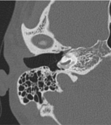
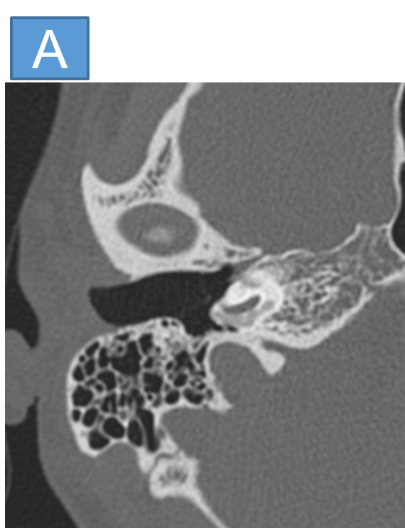
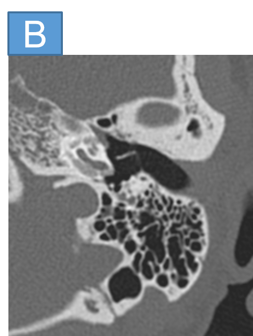
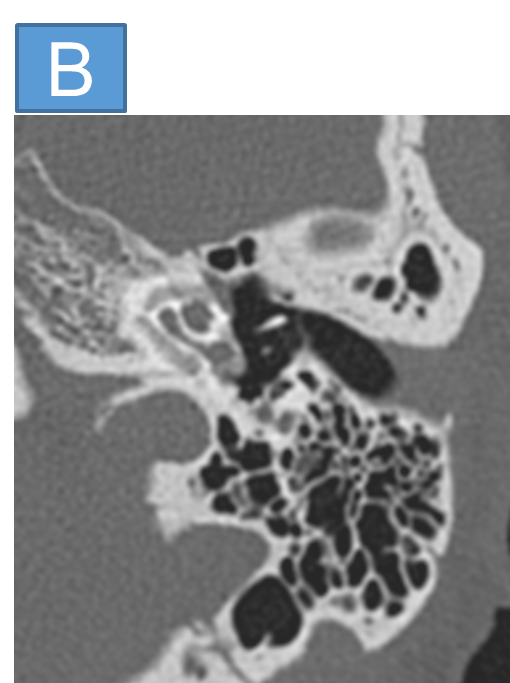
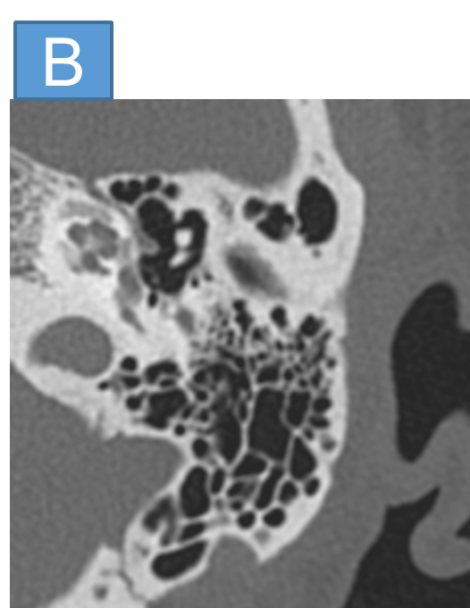
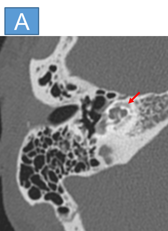
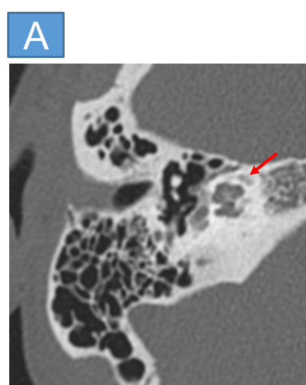
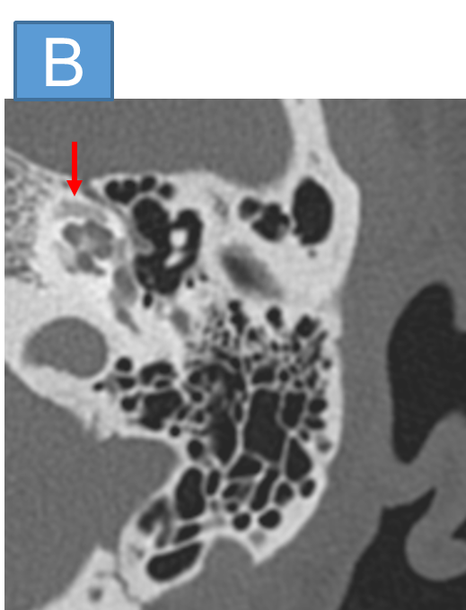
- CT TEMPORAL BONE (RIGHT SIDE)
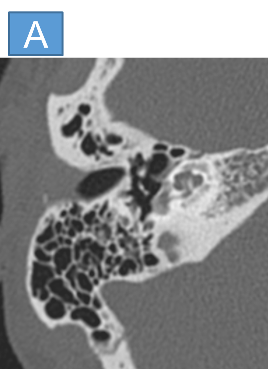
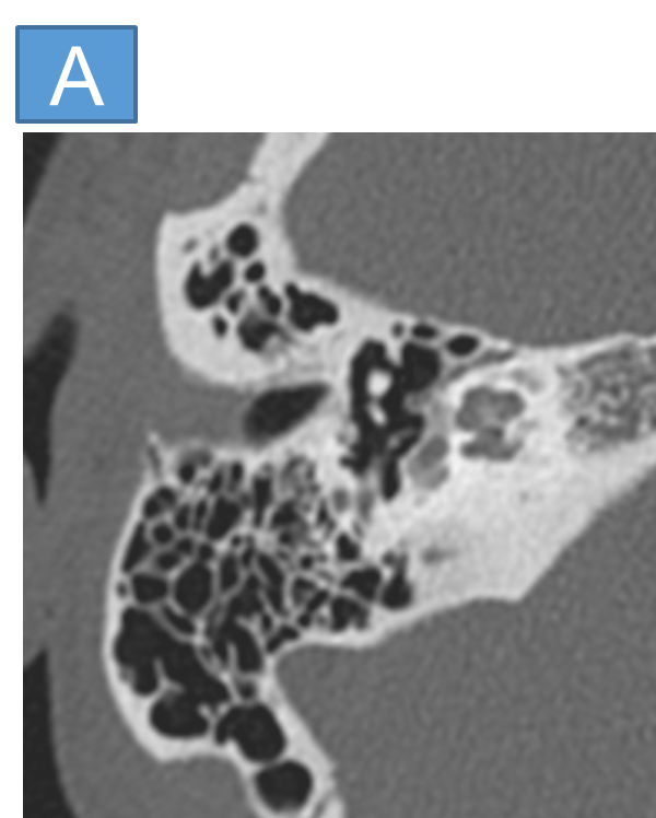
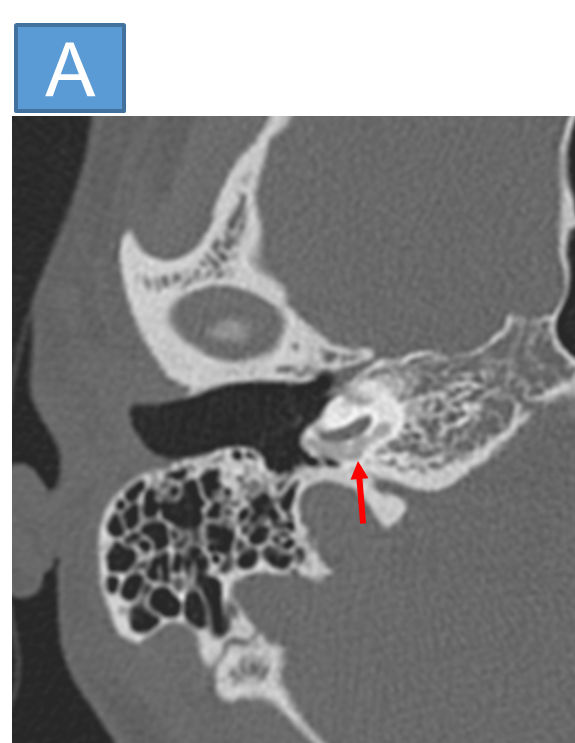
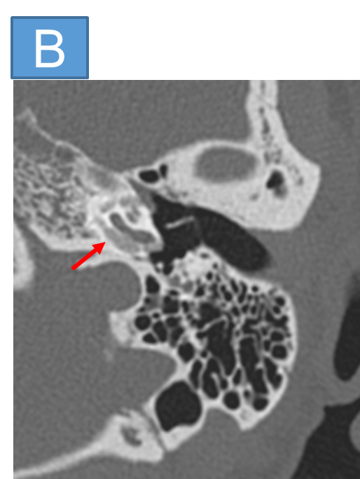
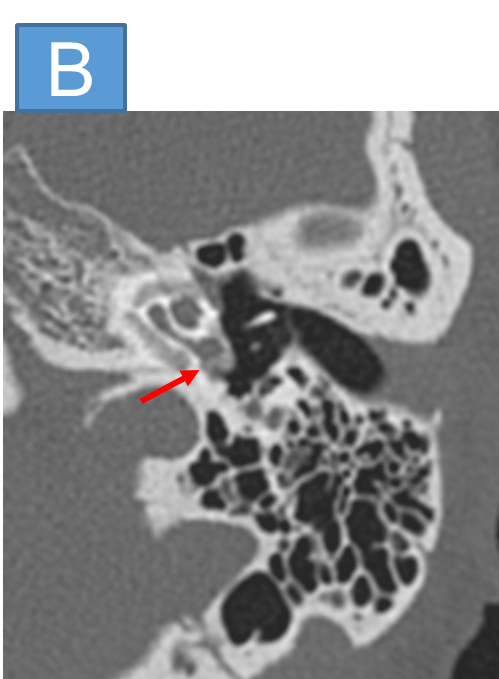
- CT TEMPORAL BONE (LEFT SIDE)
- Confluent arc like hypodense plaques are seen in the fissula ante fenestram with demineralisation around the basal, middle and apical turns of cochlea.
- There is also partial demineralisation around the around the round window niche which appears obliterated.
- There is also partial demineralisation around the basal turn of cochlea with involvement of the round window niche.
DIAGNOSIS:
- Bilateral otosclerosis - Grade 3 (Fenestral and retrofenestral type).
DISCUSSION:
- Otosclerosis is an otodystrophy of the otic capsule and is a cause of conductive, mixed or sensorineural hearing loss in the 2nd to 4th decades of life.
- It is an autosomal dominant otodystrophy of the otic capsule.
- It is also called ‘otospongiosis’ as it is characterised by replacement of the normal ivory-like enchondral bone by spongy vascular bone.
- The decalcified foci tend to recalcify, becoming less vascular and more solid.
- CLINICAL PRESENTATION: Patients typically present in the 2nd- 4th decades of life with conductive hearing loss (CHL), sensorineural hearing loss (SNHL) or mixed hearing loss (MHL) and/or tinnitus.
Otosclerosis is categorised into two types:
1) fenestral (stapedial): ~80
- involves the oval window and the stapes footplate.
- hearing loss is often conductive, due to stapes thickening and fixation.
2) retrofenestral (cochlear): ~20%
- cochlear involvement with demineralization of the cochlear capsule.
- hearing loss is often sensorineural, but the mechanism by which this occurs is uncertain.
Pathology and clinical findings:
- The more common fenestral type of otosclerosis involves the lateral wall of the bony labyrinth.
- Histologically, demineralised foci of spongy new bone typically occur in the region of the embryonic fissula ante fenestram, which is a cleft of fibrocartilagenous tissue between the inner and middle ear, just anterior to the oval window.
- Bilateral involvement is common.
- The promontory, round window niche and tympanic segment of the facial nerve canal can also be involved.
- The disease gradually extends to involve the entire footplate of the stapes and may subsequently involve the cochlea.
- Heaped-up bony plaques formed in the healing phase typically cause narrowing of the oval and round windows.
- Otosclerosis can sometimes present as isolated round window involvement without pericochlear or oval window involvement
CT grading system (Symons and Fanning):
grade 1
- solely fenestral, either spongiotic or sclerotic lesions, evident as a thickened stapes footplate, and/or decalcified, narrowed or enlarged round or oval windows
grade 2
- patchy localized cochlear disease (with or without fenestral involvement)
- grade 2A: basal cochlear turn involvement
- grade 2B: middle / apical turns involvement
- grade 2C: both the basal turn and the middle / apical turns involvement
grade 3
- diffuse confluent cochlear involvement of the otic capsule (with or without fenestral involvement)
Management:
- A stapedectomy with stapes prosthesis is the treatment of choice for fenestral otosclerosis.
References:
- Purohit, B., Hermans, R. & Op de beeck, K. Imaging in otosclerosis: A pictorial review. Insights Imaging 5, 245–252 (2014).
- https://doi?org/10?1007/s13244-014-0313-9
- T.C. Lee, R.I. Aviv, J.M. Chen, J.M. Nedzelski, A.J. Fox and S.P. Symons.
- American Journal of Neuroradiology August 2009, 30 (7) 1435-1439; DOI: https://doi.org/10.3174/ajnr.A1558
Dr ANITA NAGADI
Consultant Radiologist
Manipal Hospital, Yeshwanthpur, Bengaluru.
Dr NEHA SATHYANARAYANA
Radiology resident
Manipal Hospital, Yeshwanthpur, Bengaluru.

