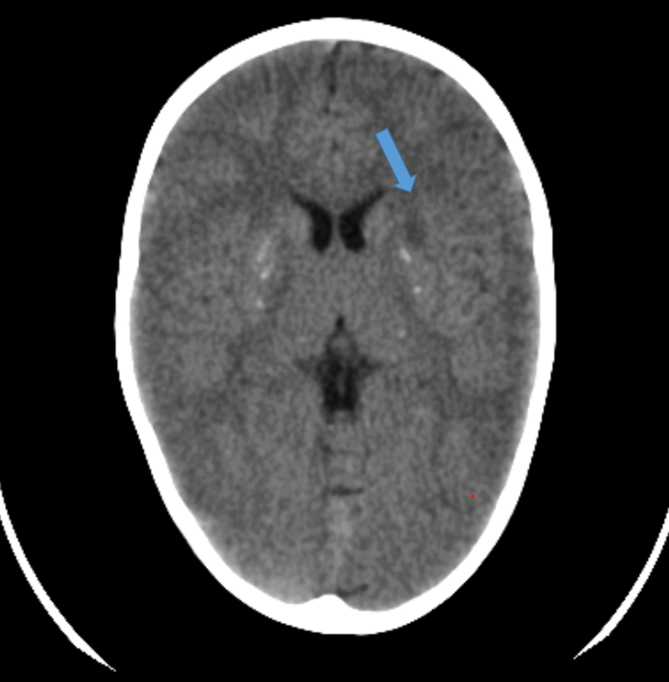
3 year old male presented with complaints of history of fall with right sided weakness
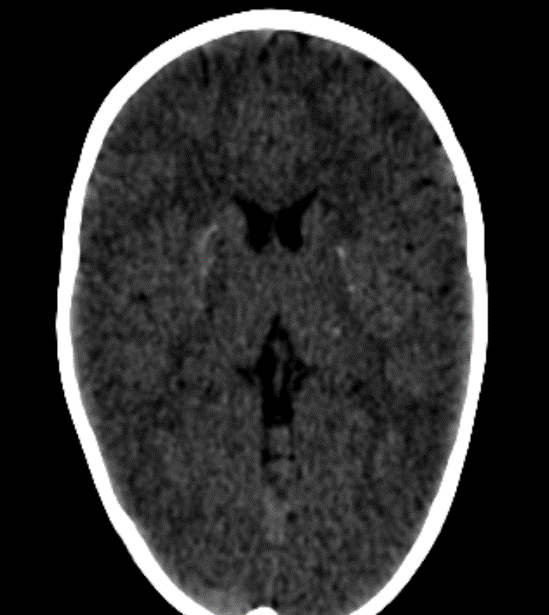
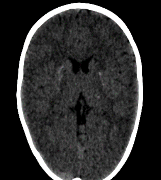
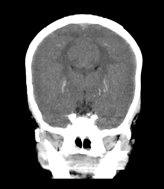
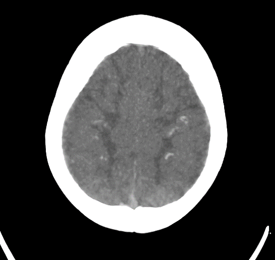
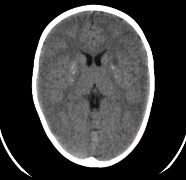
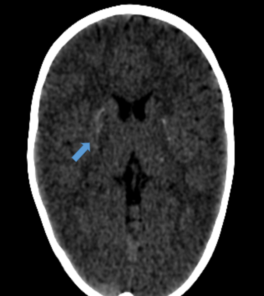
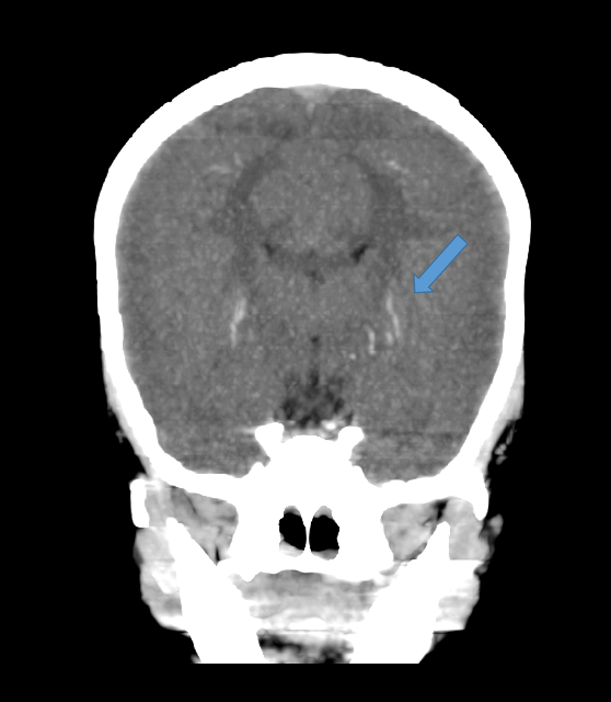
1. Axial and Coronal CT images
Shows curvilinear calcification in bilateral basal ganglia
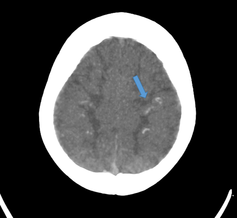
2. Image 1 shows a few linear calcifications in the subcortical white matter of the bilateral frontal lobe
Image 2- Acute infarct in left gangliocapsular region
DIAGNOSIS:
Mineralizing angiopathy with acute infarct
DISCUSSION:
Basal ganglia stroke has been reported to occur in infants in association with minor falls. Recently a distinct clinical-radiological entity termed ‘mineralizing angiopathy’ as described
Mineralizing angiopathy of lenticulostriate arteries is a common cause of subcortical strokes after minor trauma in infants.
CLINICAL PRESENTATION:
- occurrence in previously healthy infants aged 6 to 24 months
- rapid onset of neurological deficits (usually hemiparesis) minutes to hours after a minor fall
- appearance of hemidystonia on the affected side in a significant proportion of individuals, typically between day 2 and 4 after the onset of illness, subsiding within 24 hours
- In follow-up CT, mostly there maybe reduction in the intensity of mineralization
RADIOGRAPHIC FEATURES:
- Brain CT demonstrates characteristic, sharply marginated, linear hyperdense lesions coursing vertically through the inferior aspect of the lentiform nuclei in all infants.
- On coronal and sagittal reconstructions, the distribution of these linear lesions are similar to that described for lenticulostriate arteries
- These lesions had attenuation values between 58 and 90 HU, representing mineralization. The number of such mineralized vessels ranged from one to five on each side
- Infarcts in basal ganglia
CT is diagnostic of the pathology rather than MRI
Conclusion
Whenever basal ganglia stroke is seen in a paediatric age group particularly after history of trauma , look out for linear calcification in bilateral basal ganglia to rule out mineralising angiopathy
As these calcifications are difficult to visualise in MRI , its better to do a correlative CT to rule out the possibility
References :
- Lingappa L, Varma RD, Siddaiahgari S, Konanki R. Mineralizing angiopathy with infantile basal ganglia stroke after minor trauma. Dev Med Child Neurol. 2014;56:78–84. [PubMed] [Google Scholar]
- Ivanov I, Zlatareva D, Pacheva I, Panova M. Does lenticulostriate vasculopathy predispose to ischemic brain infarct? A case report. J Clin Ultrasound 2012; 40: 607–10
- Ahn JY, Han IB, Chung YS, Yoon PH, Kim SH. Posttraumatic infarction in the territory supplied by the lateral lenticulostriate artery after minor head injury. Childs Nerv Syst 2006; 22: 1493–6.
- Makhoul IR, Eisenstein I, Sujov P, et al. Neonatal lenticulostriate vasculopathy: further characterization. Arch Dis Child Fetal Neonatal Ed 2003; 88: F410–14
Dr. SRIRAM PATWARI
Senior Consultant Radiologist
Manipal Hospital, Yeshwanthpur, Bengaluru.
Dr. SHARNITHA JOHNSON
Fellow in Radiology
Manipal Hospital, Yeshwanthpur, Bengaluru.

