21-year-old male patient with a history of trauma to the knee while playing football 1yr back
- 21 yr old male patient with history of trauma to the knee while playing football 1yr back.
- Now c/o instability and pain.
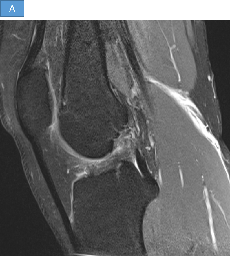
- A. Sagittal PDFS image shows Complete Tear of the Anterior Cruciate Ligament. No marrow oedema/contusions owing to old injury.
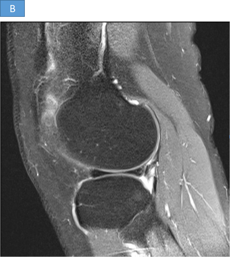
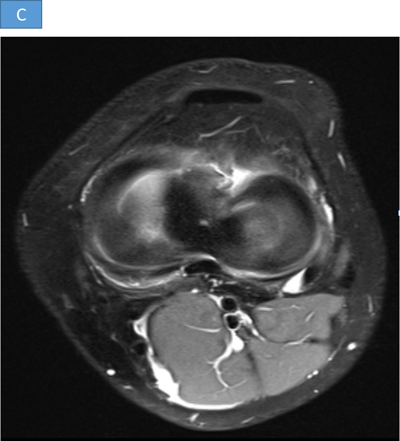
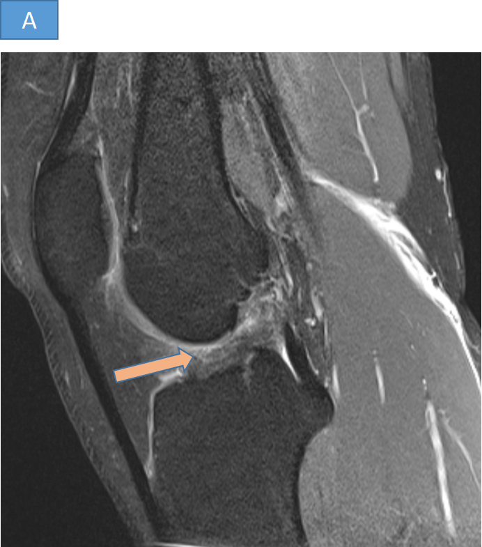
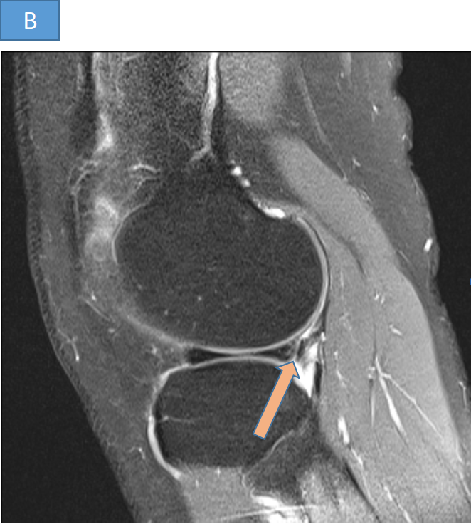
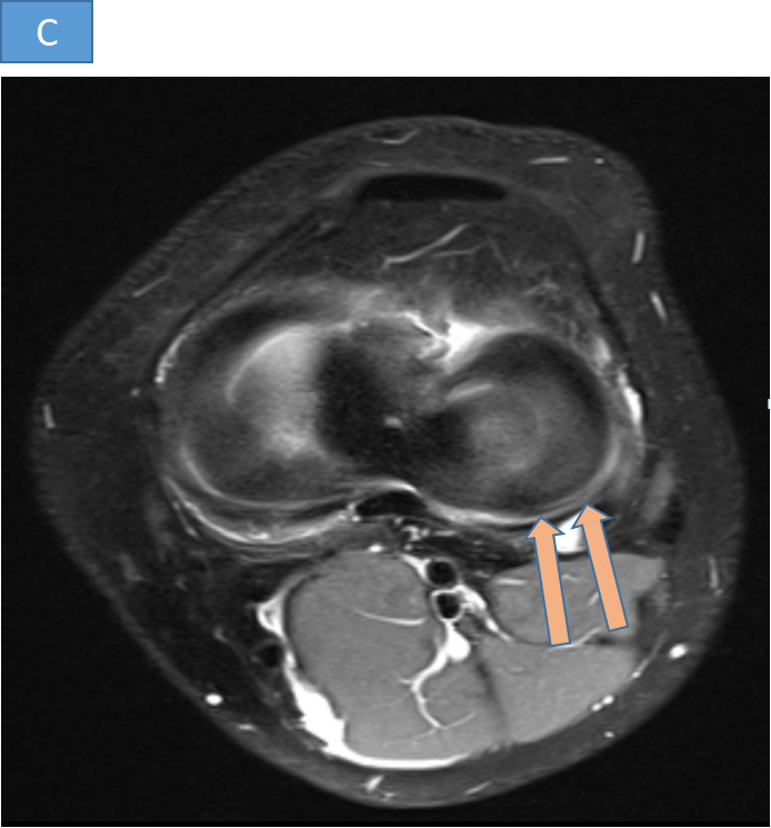
- B and C. Sagittal and Axial PDFS images show Longitudinal Vertical Tear of the Posterior Horn of the Lateral Meniscus along the Wrisberg Ligament extending laterally.
DIAGNOSIS:
- WRISBERG RIP TEAR OF LATERAL MENISCUS IN THE BACKGROUND OF CHRONIC COMPLETE ACL TEAR.
DISCUSSION:
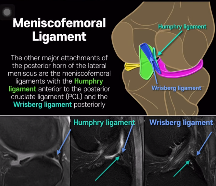
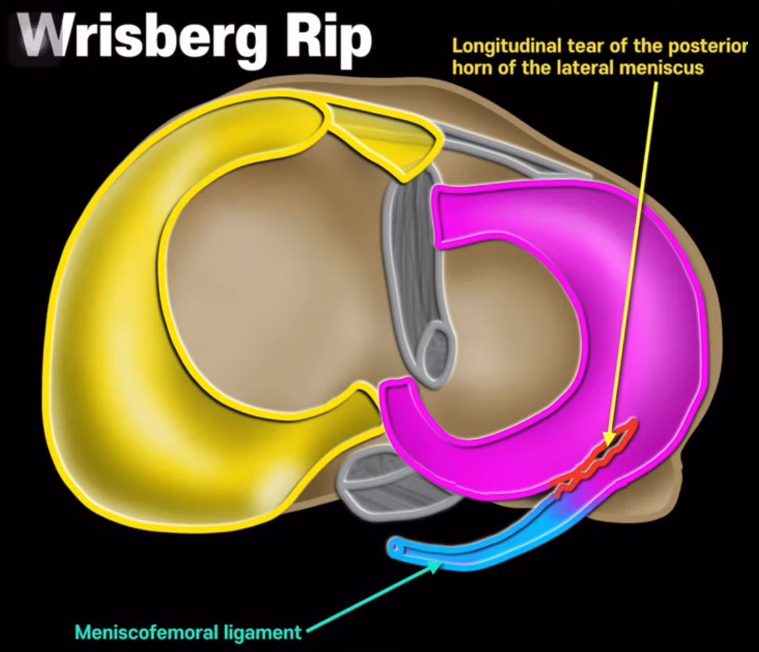
ANATOMY OF MENISCOFEMORAL LIGAMENTS
- Meniscofemoral ligament (MFL) arises from the posterior horn of the lateral meniscus and passes to attach to the lateral aspect of the medial femoral condyle.
- It splits into two bands at the posterior cruciate ligament (PCL), which are named in relation to the Posterior cruciate ligament.
1. Anterior Meniscofemoral Ligament: Ligament of Humphrey.
2. Posterior Meniscofemoral ligament: Ligament of Wrisberg.
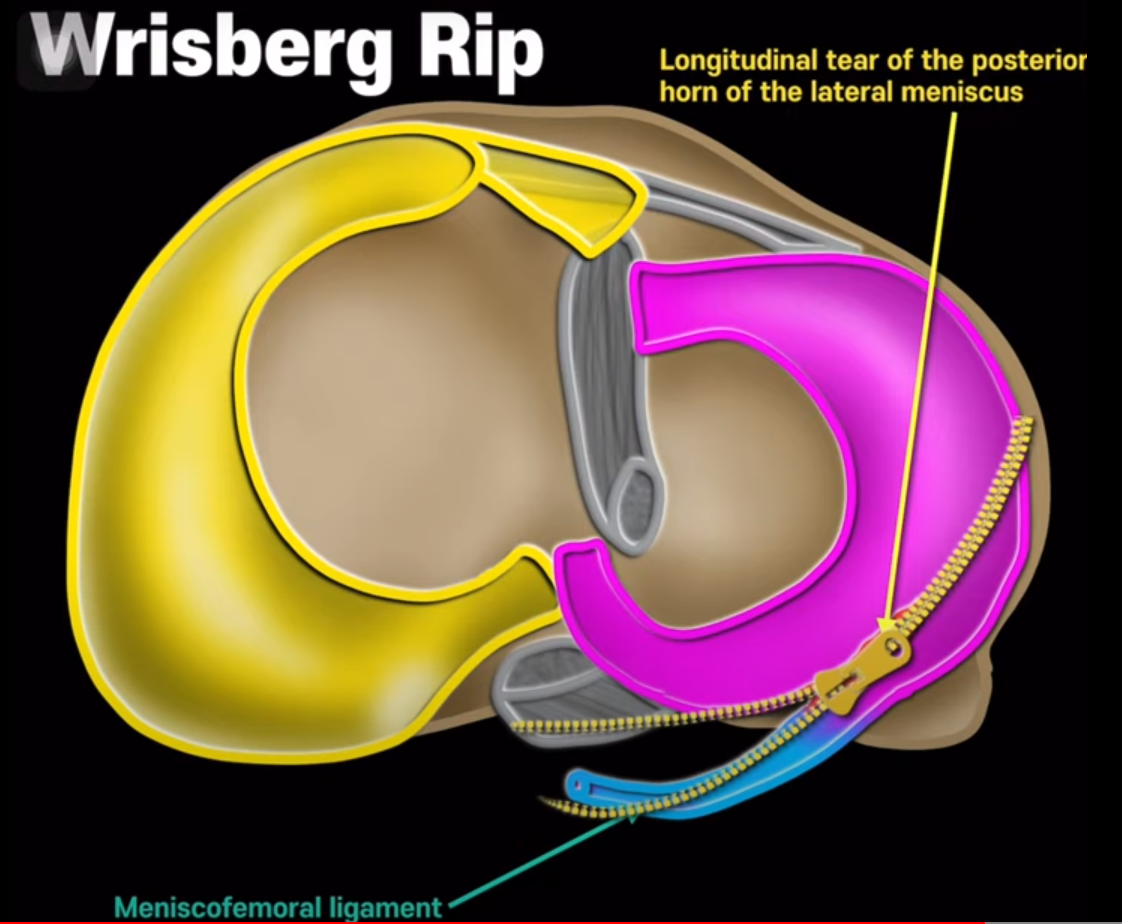
WRISBERG RIP TEAR
- DEFINITION: Subset of longitudinal tears of the posterior horn of the lateral meniscus originates at the junction of the posterior horn of the lateral meniscus and the Posterior Menisco-Femoral Ligament (Ligament of Wrisberg).
- ASSOCIATION: Always associated with ACL tear.
- MECHANISM OF INJURY: During an ACL disruption the tibia typically translates anteriorly relative to the distal femur. The Ligament of Wrisberg is attached proximally to the lateral aspect of the medial femoral condyle and distally to the posterior horn of the lateral meniscus. With ACL disruption, as the tibia moves anteriorly relative to the femur, the Ligament of Wrisberg thus causes traction on the peripheral rim of the posterior horn of the lateral meniscus. As a result, one may suffer a “zip”tear through the posterior horn of the lateral meniscus, originating at the Wrisberg attachment. Because of the unique mechanism of injury involved , this is termed as “Wrisberg Rip” tear.
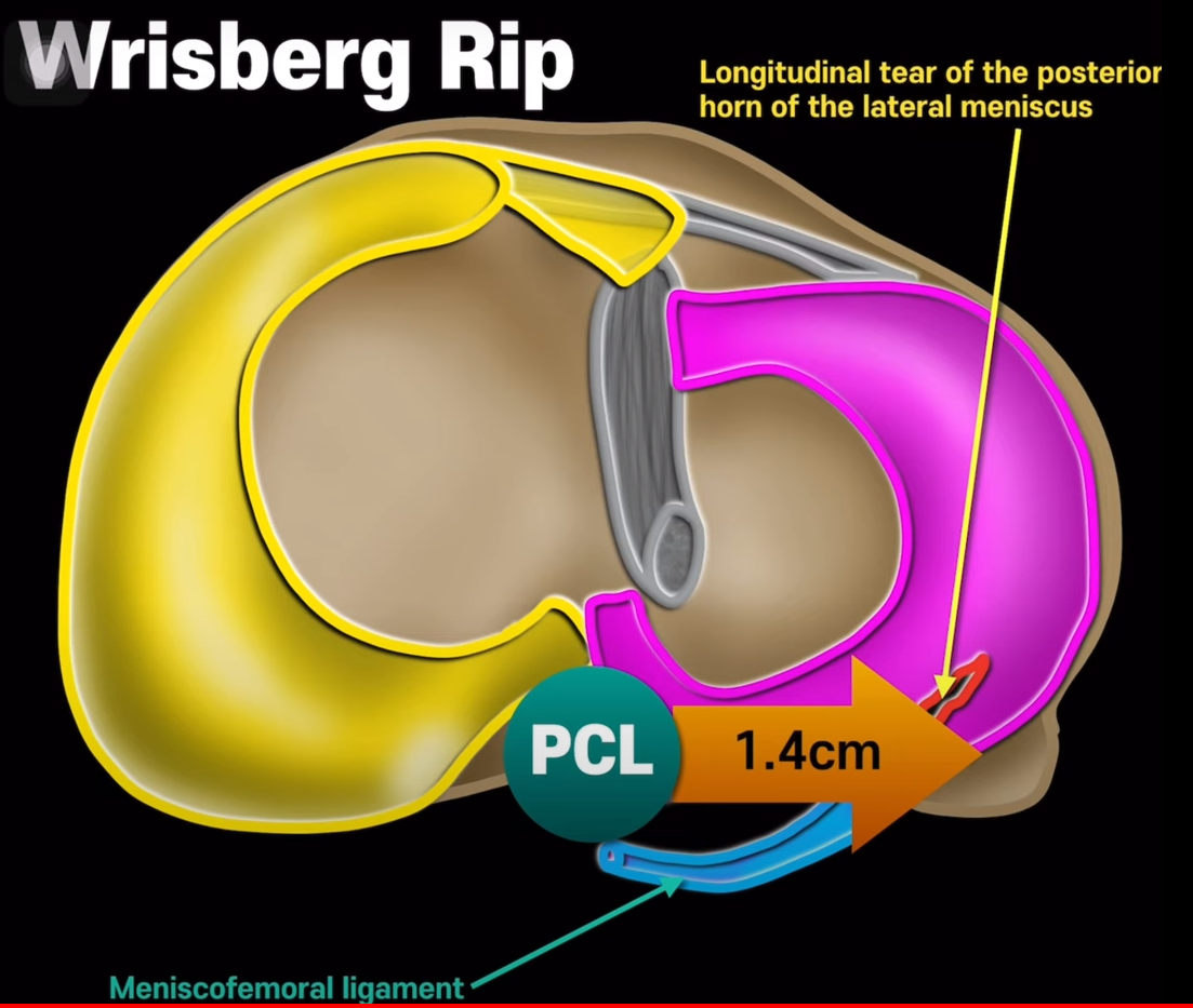
DIAGNOSIS
- The attachment site of the Ligament of Wrisberg lies approximately 14 mm laterally from the lateral edge of the Posterior cruciate ligament.
- Any cleft extending farther is suspicious for a tear.
DIFFERENTIAL DIAGNOSIS
WRISBERG PSEUDOTEAR
- Differentiation of wrisberg tear from the normal pseudo-tear is made by lateral extension of the cleft between the meniscus and meniscofemoral ligament by 14mm or more lateral to the lateral margin of the PCL.
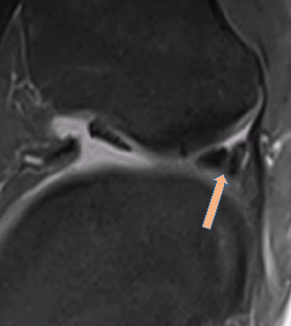
- Sagittal PDFS image showing Normal Cleft or Wrisberg Pseudotear.
IMPORTANCE
This tear type is important for two reasons.
- Tears at the central portion of the posterior horn laterally are among the most frequently missed.
- Tear pattern may be used as a secondary sign of ACL disruption as it is only associated with ACL tears.
REFERENCES
- P?kala PA, ?azarz DP, Rosa MA, P?kala JR, Baginski A, Gobbi A, Wojciechowski W, Tomaszewski KA, LaPrade RF. Clinical anatomy of the posterior meniscofemoral ligament of Wrisberg: an original MRI study, meta-analysis, and systematic review. Orthopaedic Journal of Sports Medicine. 2021 Feb 22;9(2):2325967120973195.
- Tomsan H, Gorbachova T, Fritz RC, Abrams GD, Sherman SL, Shea KG, Boutin RD. Knee MRI: meniscus roots, ramps, repairs, and repercussions. RadioGraphics. 2023 Jun 29;43(7):e220208.
- Lecouvet F, Van Haver T, Acid S, Perlepe V, Kirchgesner T, Berg BV, Triqueneaux P, Denis ML, Thienpont E, Malghem J. Magnetic resonance imaging (MRI) of the knee: Identification of difficult-to-diagnose meniscal lesions. Diagnostic and interventional imaging. 2018 Feb 1;99(2):55-64.
- Saad SS, Gorbachova T, Saing M. Meniscal tears: scanned, scoped, and sculpted: resident and fellow education feature. Radiographics. 2015 Jul;35(4):1138-9.
- Stensby JD, Pringle LC, Crim J. MRI of the Meniscus. Clinics in Sports Medicine. 2021 Oct 1;40(4):641-55.
- Mohankumar R, White LM, Naraghi A. Pitfalls and pearls in MRI of the knee. American Journal of Roentgenology. 2014 Sep;203(3):516-30.
Dr. DEEPTI H V
CONSULTANT RADIOLOGIST
MANIPAL HOSPITAL , YESHWANTHPUR, BENGALURU
Dr. JOMON SUNNY
FELLOW IN CROSS SECTIONAL IMAGING
MANIPAL HOSPITAL , YESHWANTHPUR, BENGALURU