
17 years male. History of abdominal discomfort and long-standing constipation.
History
17 years male.
History of abdominal discomfort and long-standing constipation.
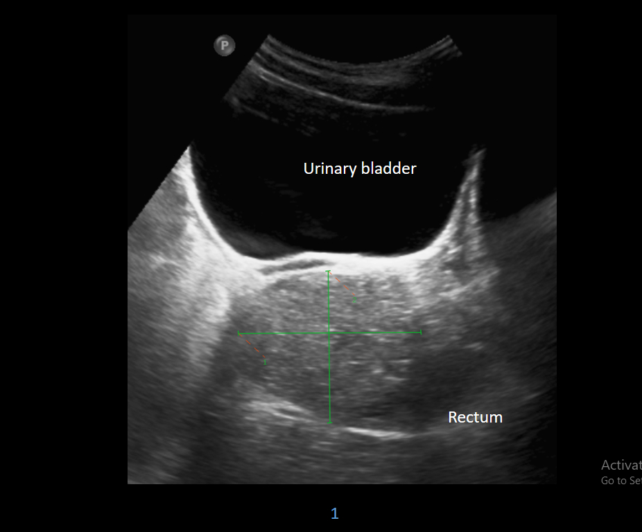
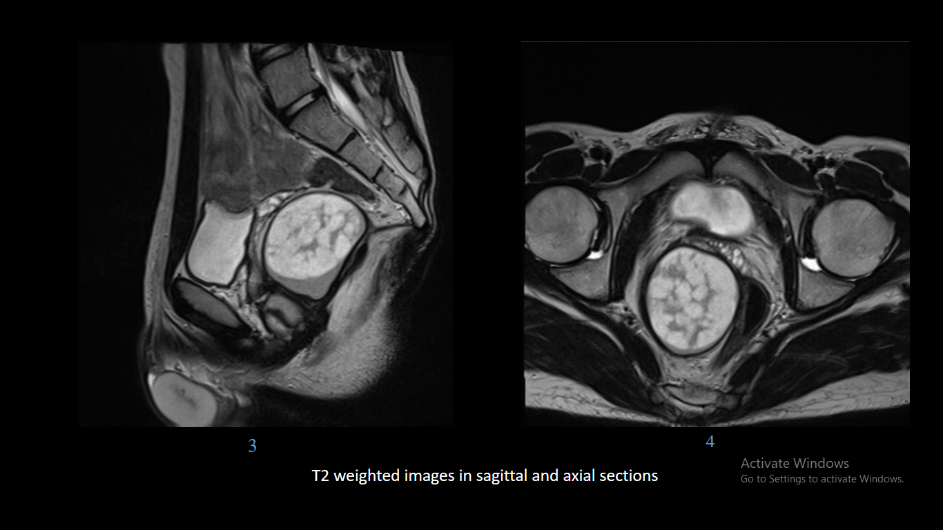
Ultrasound demonstrates a hyperechoic lesion in the pelvis on the right side abutting
the rectum.
Diagnosis
Pre sacral epidermoid cyst
Laparotomy with complete surgical excision of the cyst was performed. The histopathology confirmed the diagnosis of epidermoid cyst.
Discussion
Epidermoid cysts:
- Represent proliferation of squamous epithelium within a confined space.
- Either found incidentally or present as a firm non-tender lump.
- If they rupture a local inflammatory response to the necrotic debris released can mimic infection.
- Although they can be found anywhere, they are typically located on the scalp, face, neck, trunk, and back.
- Rarely epidermal cysts can undergo malignant degeneration with squamous cell carcinoma.
Pathology:
- They are thought to occur as a result of:
- Traumatic/surgical implantation.
- Occlusion of the pilosebaceous unit.
- Congenital rests of cells.
- Human papillomavirus type 57 or 60 infections implicated palm plantar epidermoid cysts.
Pre Sacral Epidermoid Cysts
- Developmental cystic lesions arising in the presacral location include dermoid cysts, epidermoid cysts, chordomas, adrenal rest tumors, anterior sacral meningoceles, cystic hamartomas, tailgut or rectal duplication cyst.
- Natural history of epidermoid cysts arising in the presacral region is elusive. Because of their unusual location and slow growth, they have a tendency to remain asymptomatic.
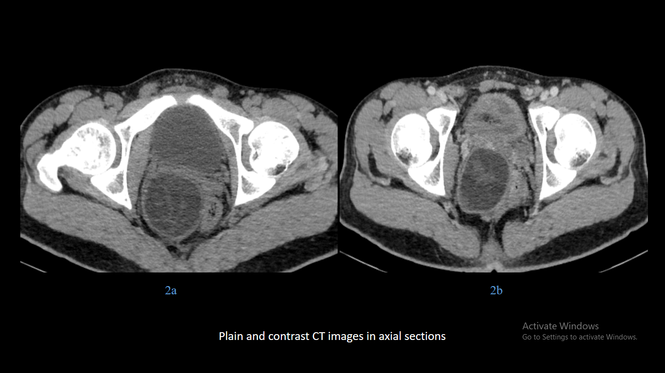
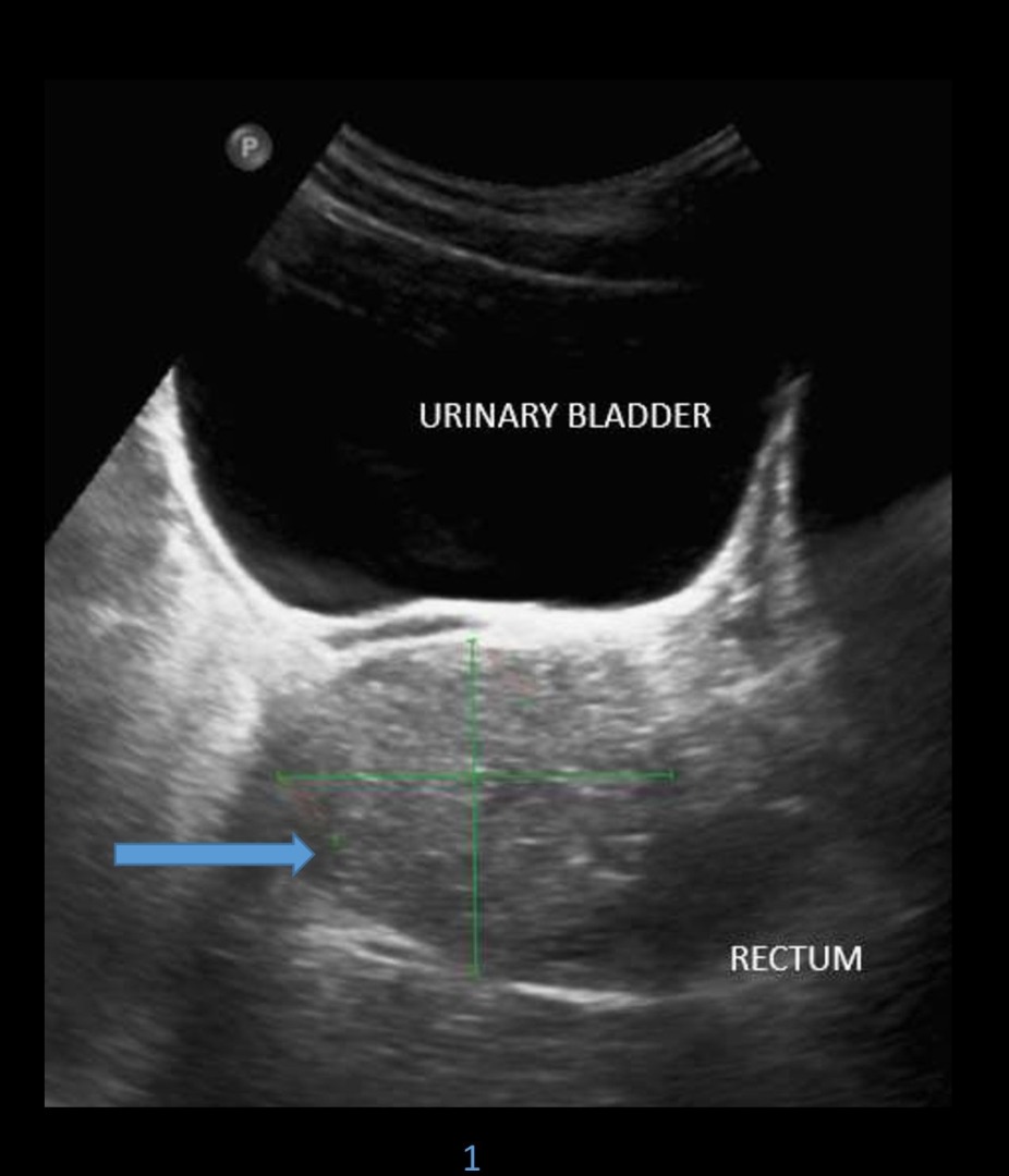
- Difficult to characterize on ultrasound and computed tomography.
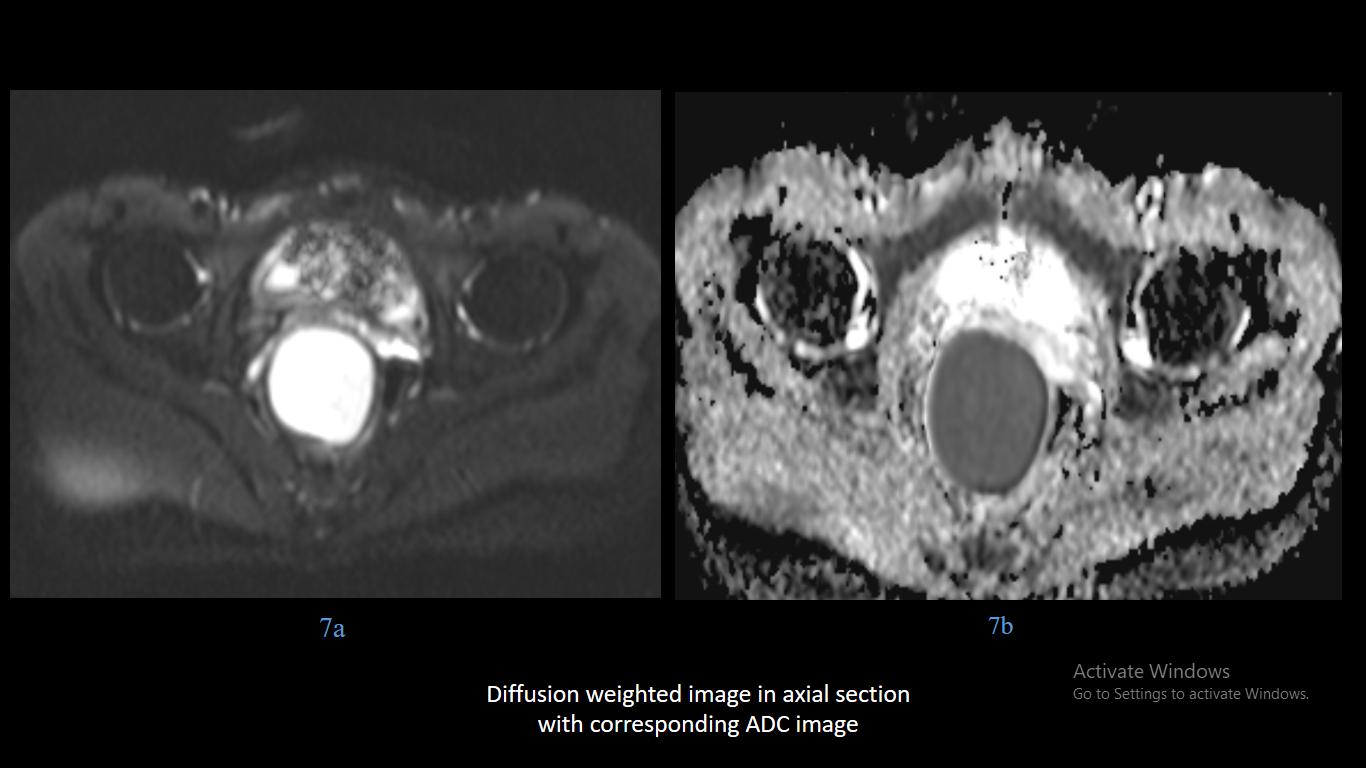
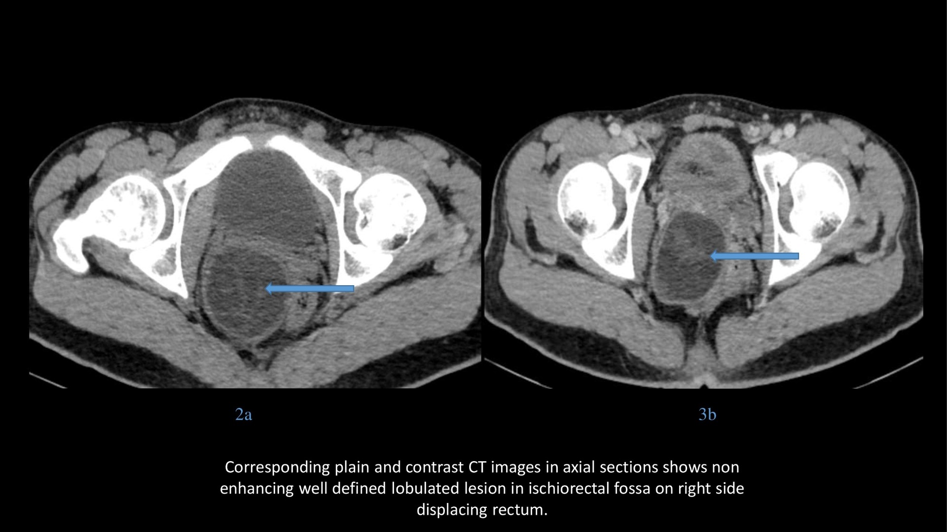
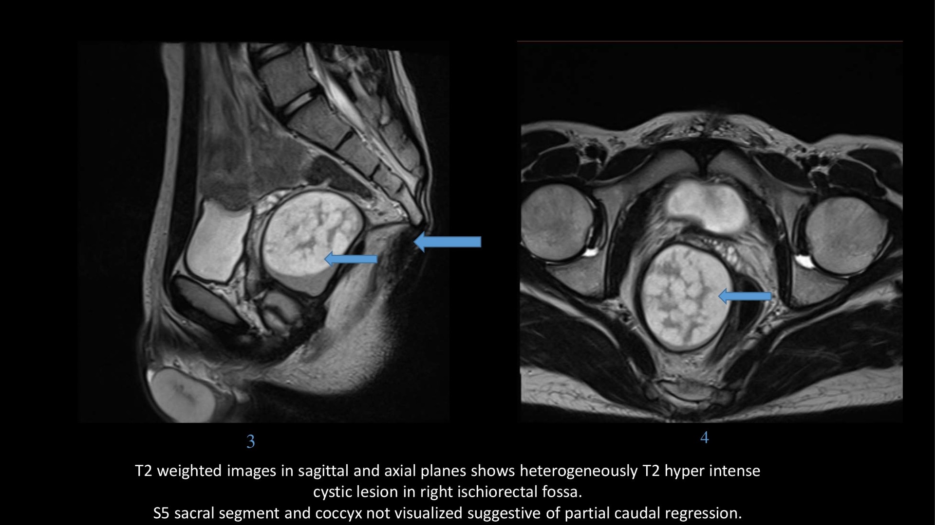
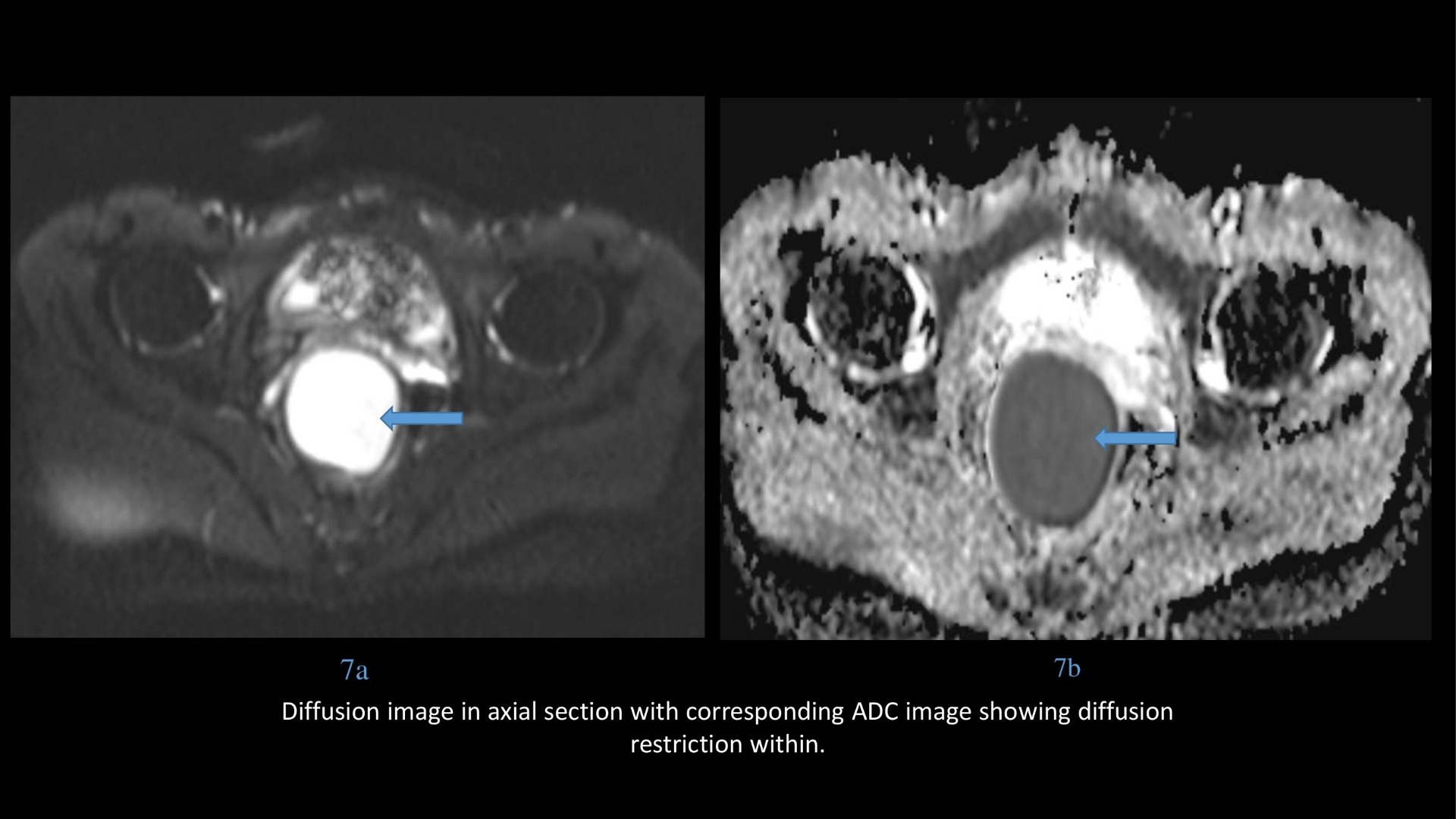
- MRI with the use of diffusion weighted imaging allows for definite preoperative diagnosis.
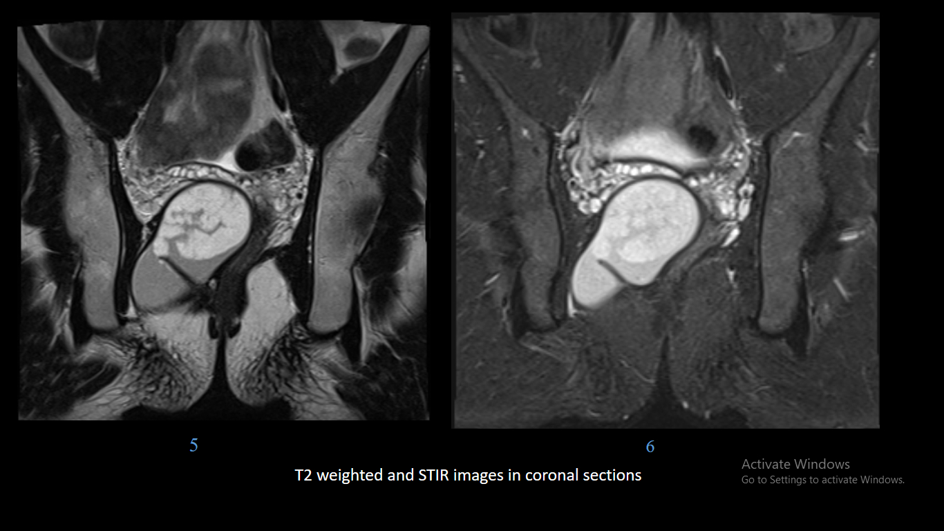
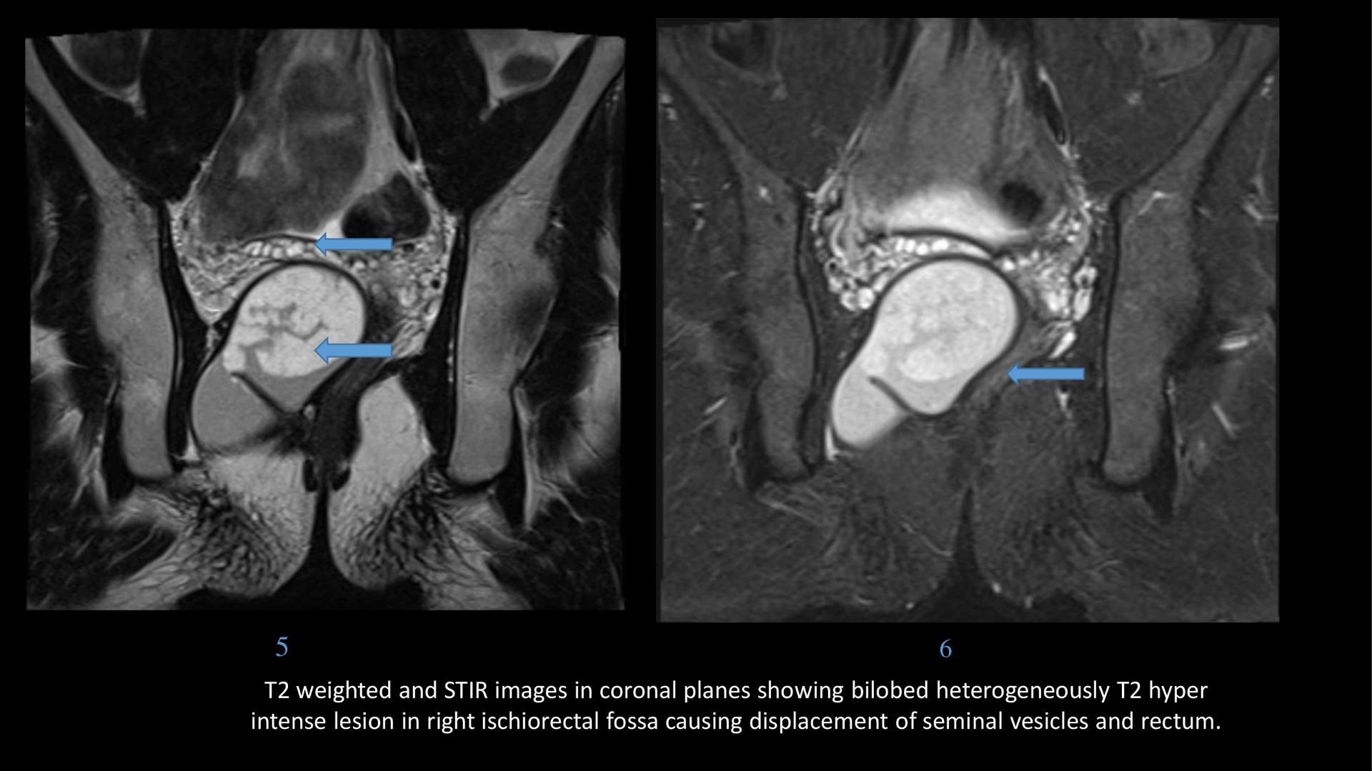
- Epidermoid cysts appear as T1 hypointense, T2 hyperintense masses which show diffusion restriction, wherever they are present within the body.
- T2 hypointense foci may be seen within the lesion because of presence of keratin.
- Another feature described in pre sacral epidermoid is differential restriction in contents of the cyst.
- MRI also helps differentiating between any bony, spinal canal or meningeal involvement, or signs of malignant degeneration.
- Heterogeneous signal intensity on T2, irregular thickened walls, solid component, and presence of enhancement are concerning for malignant change.
- A close differential of an epidermoid cyst on basis of diffusion restriction is a retrorectal pyogenic abscess. However abscess demonstrates presence of a thickened enhancing rim, intracavity fluid debris level or air specks; surrounding fat may show inflammatory changes with thickening of rectal wall.
Summary
- Presacral epidermoid cysts are unique in that they are the only developmental lesions that show diffusion restriction.
- MRI with diffusion weighted imaging allows for accurate differentiation of pre sacral lesions with good confidence thereby obviating the need of biopsy before surgery.
References
- U. Wollina, D. Langner, G. Tchernev, K. França, T. Lotti, Epidermoid cysts – A wide spectrum of clinical presentation and successful treatment by surgery: a retrospective 10-year analysis and literature review, Open Access Maced. J. Med. Sci. 6 (1) (2018) 28–30, https://doi.org/10.3889/oamjms.2018.027.
- H. Curtin, P. Som, Head and Neck Imaging, 4th ed., Mosby, 2002, pp. 2173–2183, https://doi.org/10.1016/S0720-048X(03)00181-5.
- Yang BL, Gu YF, Shao WJ, et al.: Retrorectal tumors in adults: magnetic resonance imaging findings. World J Gastroenterol. 2010, 14:5822-5829. 10.3748%2Fwjg.v16.i46.5822.
- Riojas CM, Hahn CD, Johnson EK: Presacral epidermoid cyst in a male: a case report and literature review. J Surg Educ. 2010, 31:227–232.
- Yang DM, Kim HC, Lee HL, et al.: Squamous cell carcinoma arising from a presacral epidermoid cyst: CT and MR findings. Abdom Imaging. 2008, 33:498-500. 10.1007/s00261- 007-9287-0.
- Izumi K, Tsutsumi S, Hara T, et al.: Large presacral epidermoid cyst in an asymptomatic woman. Radiol Case Rep. 2017, 12:738–740. 10.1016/j.radcr.2017.07.017
Dr. Srinivas P
Cross-sectional Fellow
Manipal Hospital Radiology Group (MHRG)
Manipal Hospital, Bengaluru.
Dr. Vishwanath Joshi
Consultant Radiologist.
Manipal Hospital Radiology Group (MHRG)
Manipal Hospital, Bengaluru.
