
A 32-year-old lady with bilateral hearing loss and vertigo. No history of prior surgery
A 32-year-old lady with bilateral hearing loss and vertigo. No history of prior surgery.
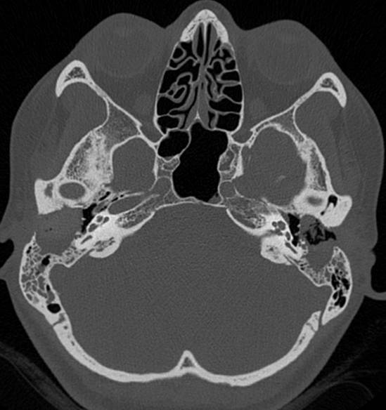
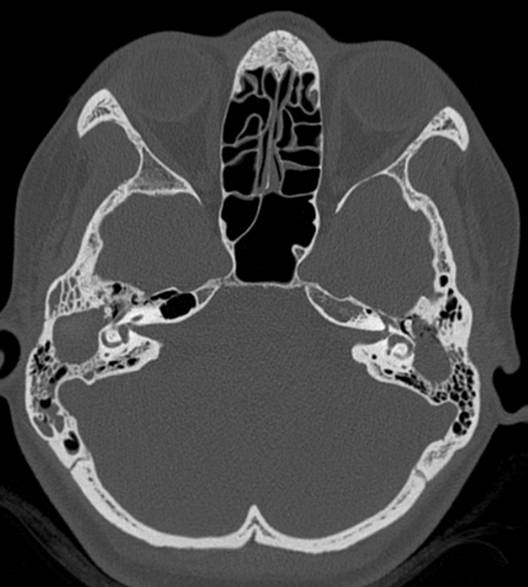

- Axial HRCT images of temporal bones demonstrate heterogenous opacification of the external auditory canals, with marked destruction of the posterior canal walls and extension of opacification into the mastoids. There is relative sparing of the middle ear clefts bilaterally. Opacification of the left epitympanum and a labyrinthine fistula is noted.
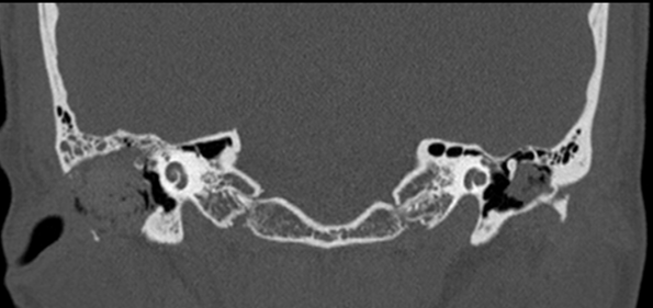
- Coronal reformats demonstrate dehiscence of the mastoid segments of the facial nerve bilaterally. There is also bony dehiscence of the anterior tympanic segment of the right facial nerve.
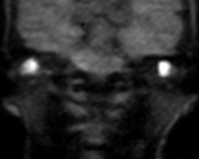
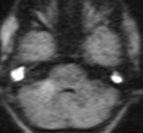
- Axial and coronal non-EPI DWI images demonstrate marked restricted diffusion within both external auditory canals corresponding to the opacification seen on CT.
Diagnosis:
Bilateral EAC cholesteatoma with left labyrinthine fistula and bilateral facial nerve canal dehiscence.
Discussion:
Cholesteatoma is an inflammatory lesion of the temporal bone that uncommonly involves the external auditory canal
EAC cholesteatoma is usually seen in the older population.
Types:
- Primary: Idiopathic. Secondary to loss of normal migration in aging epithelium of the canal wall
- Secondary: Post-operative, post-inflammatory, post-traumatic, radiation-induced. It can be associated with congenital atresia of the external auditory canal.
Imaging:
- It is associated with smooth or irregular bony erosion and can be difficult to distinguish from SCC of external auditory canal. Otoscopy and biopsy should be performed.
- EAC cholesteatoma is often more extensive than that suggested by the clinical findings.
- Important landmarks in the petrous temporal bone to be evaluated for their integrity: facial nerve canal, tegmen tympani (roof of the middle ear cavity), semicircular canals, and mastoid air cells
Differential diagnosis:
- Keratosis obturans: Present with acute, severe otalgia and conductive hearing loss. The soft tissue in EAC, with bony remodeling. The absence of erosions helps distinguish it from EAC cholesteatoma.
- Necrotizing otitis externa: Necrotising infection of the external canal in elderly and diabetics. Associated with soft tissue inflammatory changes, which can extend into the mastoid, skull base, and neck.
- Malignancy (rare):
- Squamous cell carcinoma: Metastasize early.
- Basal cell carcinoma: Locally aggressive and rarely metastasize.
- Minor salivary gland tumors
Dr. Akshay K,
Radiology Resident,
Manipal Hospitals Radiology Group.
Dr. Anita Nagadi,
MD, MRCPCH, FRCR, CCT (UK)
Lead Head & Neck, Oncology
Senior Consultant Radiologist
Manipal Hospitals Radiology Group.