
A 58-year old gentleman presenting with constipation since 10 days
A 58-year-old gentleman presented with constipation for 10 days
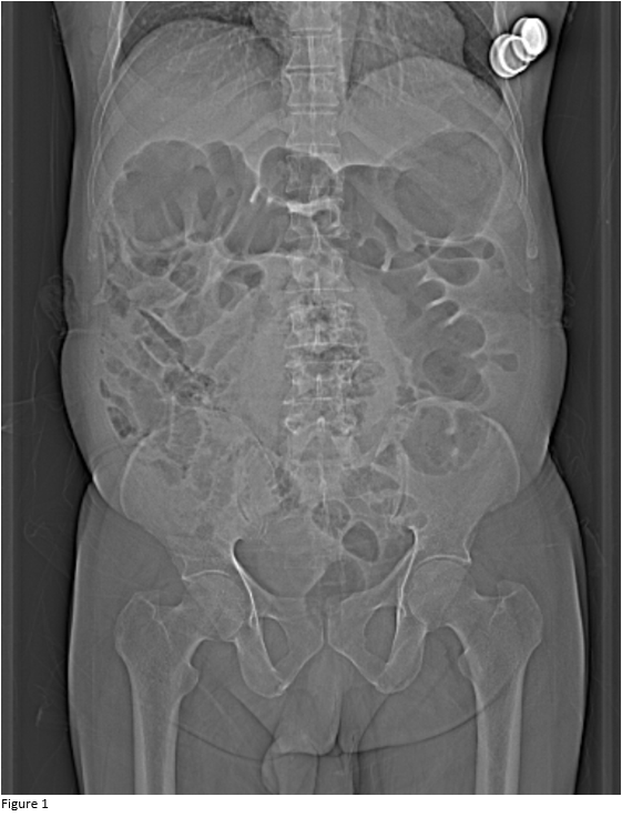
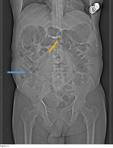
- Figure 1: Coronal sonogram demonstrates small bowel loops lying on the right side (blue arrow) with a hint of medial deviation of ascending colon (yellow arrow) and an ill-defined outline of the caecum.
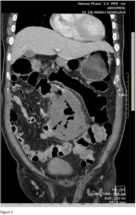
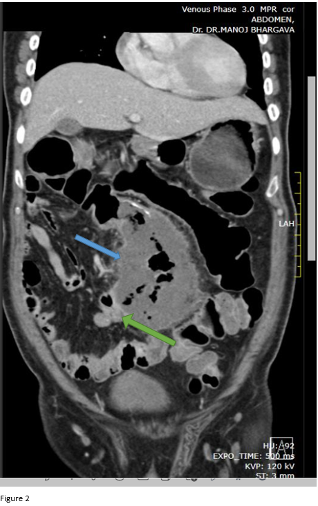
- Figure 2: Coronal oblique contrast-enhanced CT abdomen demonstrates lateral ileocaecal junction (green arrow). Ascending colon is medially deviated with dilated caecum lying in midline (blue arrow).
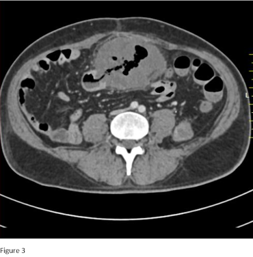
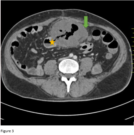
- Figure 3: Axial contrast-enhanced CT abdomen demonstrates dilated bowel loop (caecum) in the midline with the poor enhancement of left-sided wall and surrounding fat stranding (green arrow) signifying wall ischemia/ necrosis; ileocaecal junction again is seen facing laterally (yellow star).
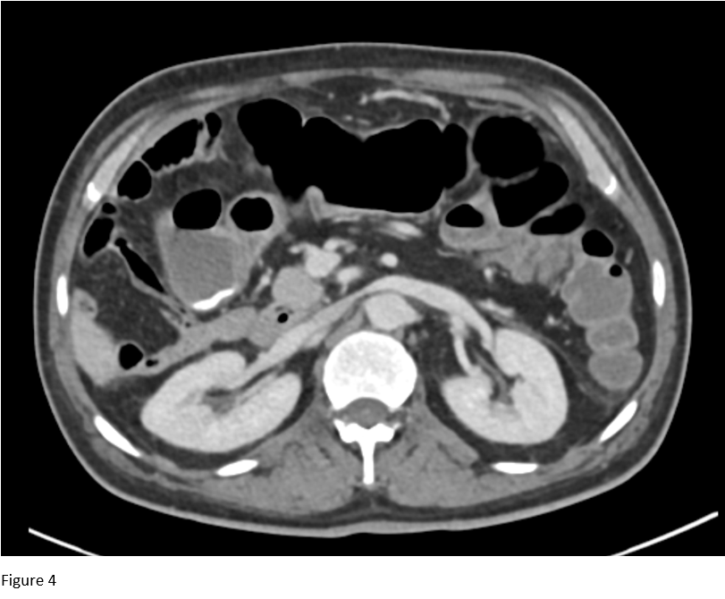
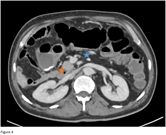
- Figure 4: Axial contrast-enhanced CT abdomen at a superior level demonstrates the absence of crossing over of duodenum across the midline, in its usual position (blue star) and abnormally low and right-sided placed duodenojejunal junction (yellow star).
DIAGNOSIS:
Malrotation (Non-rotation) of the bowel with midline gangrenous caecum possibly Torsion-detorsion volvulus of cecum.
DISCUSSION:
- Caecal volvulus usually affects patients aged 30- 60 years.
- Clinical features may vary from acute colicky pain to mild symptoms like distention, constipation nausea and vomiting.
- It usually consists of bowel twist involving the caecum, ascending colon and sometimes the terminal ileum, often resulting in closed loop obstruction.
- However caecal bascule is a subtype (Type III) of volvulus, where there is no obvious twist; but caecum comes to lie in midline.
- In the presence of an abnormal lying ceacum, it is important to assess bowel for malrotation / non rotation as in this study.
Imaging features of caecal bascule are:
- It is an extremely rare type of intestinal obstruction in which a mobile caecum folds upward across bands that may be congenital, post-operative, or inflammatory and obstructing it through a valvular mechanism.
- The caecum folds anteriorly without twisting and lies in an abnormal position.
- It is necessary to exclude the presence of a whirl sign, which signifies a twist and hence excludes a caecal bascule.
Differential diagnosis:
Acute Typhlitis:
- It is a necrotizing inflammatory condition of the caecum,
- Caecal wall thickening, caecal dilatation, fat stranding, ileus, and obstruction can be seen.
- WBC counts with a neutropenic pattern shall favor this diagnosis.
REFERENCES:
- Tonerini M, Pancrazi F, Lorenzi S, Pacciardi F, Ruschi F, et al. (2015) Cecal volvulus: what the radiologist needs to know. Glob Surg, 1: DOI: 10.15761/GOS.1000106.
- Ramsingh, Jason, et al. “Bascule caecal volvulus: a rare cause of intestinal obstruction.” Journal of surgical case reports vol. 2014,4 rju025. 11 Apr. 2014, doi:10.1093/jscr/rju025
- Caecal Bascule,URL : https://www.eurorad.org/case/12277,
Case contributed by:
Dr. Sushant Mittal. MD.
Cross-sectional fellow
Columbia Asia Radiology Group
Dr. Rutuparna S MD. ,
Consultant Radiologist
Columbia Asia Radiology Group
Dr. Sunita Gopalan DMRD, FRCR.
Senior Consultant Radiologist
Columbia Asia Radiology Group