Careers | +91 96069 52813 | teleradiology.india@manipalhospitals.com |     
HealthFlex
×
  • About Us
  • Our Presence
    • Uganda
    • UAE
    • Saudi Arabia
    • Qatar
    • Nigeria
    • Oman
    • Namibia
    • Kenya
    • Israel
    • Ghana
    • Bahrain
    • Kuwait
  • Services
    • Teleradiology Reporting Services
    • Radiology Education
      • Cross-Sectional Fellowship 2023-24
      • Manipal Hospitals FRCR 2B Course
    • Radiology Consulting
    • Artificial Intelligence
    • Research and Collaboration
  • Radiology Team
    • Radiologist team
    • Administrative team
  • Case of the week
  • Resources
    • Academics
    • Blog
  • Events
  • Contact

A 43-year gentleman with recurrent hemoptysis, recurrent genital and oral ulcers since 5 years.

A 43-year gentleman with recurrent hemoptysis, recurrent genital and oral ulcers since 5 years.
March 22, 2021
  • Case details
  • Legends
  • Diagnosis and Discussion

Case details

A 43-year gentleman with recurrent hemoptysis, recurrent genital and oral ulcers since 5 years.

Legends

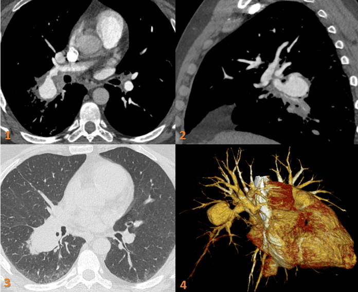
  • Fig 1 & 2. Axial and Sagittal contrast CT Pulmonary angiogram images demonstrate a large saccular aneurysm arising from the superior segmental artery of right lower lobar pulmonary artery with diffuse wall thickening – probable eccentric thrombus. There is compression of distal lower lobe segmental pulmonary arteries by the aneurysm (arrow).
  • Fig 3. Axial CT image Lung window: Perifocal ground glass opacity along the saccular aneurysm in the right lower lobe. 
  • Fig 4. VR image of pulmonary artery aneurysm.

Post coiling of pulmonary artery aneurysm.

Diagnosis and Discussion

DIAGNOSIS:

Right pulmonary artery aneurysm with partial eccentric thrombus.

DISCUSSION:

Behçet disease is a multisystemic and chronic relapsing inflammatory disorder characterized by the histopathologic finding of nonspecific vasculitis involving various sized vessels in multiple organs.

Key imaging manifestations are:

  1. Pulmonary thromboembolism.
  2. Pulmonary artery aneurysm.
  3. Sinus of Valsalva aneurysm.
  4. SVC syndrome.
  5. Intracardiac thrombosis.
  6. Mediastinal fibrosis.
  7. Intestinal ulceration (and inflammatory changes).
  8. Budd-Chiari syndrome.
  9. Cerebral venous thrombosis.
  • Identification of a vascular lesion is important because it seriously affects a patient’s prognosis. The leading cause of sudden death in patients with Behçet disease is rupture of a large aortic or arterial aneurysm
  • To improve the detection of these various radiologic findings, examinations should be well-selected and optimized in patients with Behçet disease. 

REFERENCES:

  1. Mehdipoor G, Davatchi F, Ghoreishian H, Shabestari AA. Imaging manifestations of Behcet’s disease: key considerations and major features. European journal of radiology. 2018 Jan 1;98:214-25.
  2. Chae EJ, Do KH, Seo JB, Park SH, Kang JW, Jang YM, Lee JS, Song JW, Song KS, Lee JH, Kim AY. Radiologic and clinical findings of Behçet disease: comprehensive review of multisystemic involvement. Radiographics. 2008 Sep;28(5):e31.

Case contributed by:

Dr. Deepali Saxena,
DNB, Fellowship Cardiothoracic Imaging (USA)
Lead Cardiothoracic Imaging
Manipal Hospitals Radiology Group.
Unit coordinator, Whitefield
Dr. Vivek Jirankali,
MD
Senior resident and cross-sectional fellow
Manipal Hospitals Radiology Group.

Add Comment Cancel


Contact Us

(+91) 96069 52813

teleradiology.india@manipalhospitals.com

The Annexe, No. 98/2 Rustom Bagh, Off HAL Airport Road, Bengaluru

Follow us on


Quick Links

  • Radiology Team
  • Teleradiology Reporting Services
  • Radiology Education
  • Radiology Consulting
  • Artificial Intelligence
  • Research and Collaboration
  • Case of the Week
  • Academics
  • Blog
  • Careers
  • Contact

Our Presence

  • Uganda
  • UAE
  • Saudi Arabia
  • Qatar
  • Oman
  • Nigeria
  • Namibia
  • Kenya
  • Israel
  • Ghana
  • Bahrain
  • Kuwait


Manipal Hospitals Radiology Group (MHRG) has been successfully providing tele-reporting and radpeer review services to healthcare providers across the Indian subcontinent, Indonesia, Africa, France, United Kingdom and the United States.

Read more
SITEMAP  |   DISCLAIMER  |   PRIVACY POLICY
Copyright ©2022 all rights reserved
  • +91 96069 52813 (Business Calls)

  • Email

Dr. Prakash Naik

MBBS, DMRD, FRCR

Senior Consultant Radiology – Manipal Hospitals Radiology Group

Register Now


Apply Here


Dr. Harsha Chadaga

MBBS, DMRD, DNB Radio Diagnosis,
PDCC Neuro intervention, EDiNR

Lead Neurology Imaging – Manipal Hospitals Radiology Group

  • Past President British Society of Interventional Radiology
  • Former Head of Training for Radiology in East Yorkshire
  • Couch Kerley Travelling Professorship 2012
  • Served as a Senior Examiner for the Royal College of Radiologists
Special interests: Cardiovascular Imaging
Vascular Interventional Radiology

Dr. Praveen Wali

MBBS, DMRD, DNB, FRCR, EDIR

Consultant – Radiology – Manipal Hospitals Radiology Group

Dr. Rajesh Lavakumar

MBBS, MRCS(UK), FRCR(UK), CCT(UK)

Lead – MSK Radiology – Manipal Hospitals Radiology Group

Dr. Anita Nagadi

MBBS, MD, MRCPCH (UK), FRCR (UK), CCT (UK)

Lead Head & Neck

Oncology – Manipal Hospitals Radiology Group

Dr. Raviraj Uppoor Bhat

FRCR, DNB, DMRD

Consultant Radiologist at Royal Lancaster Infirmary

UHMBT NHS Trust Special interests – Uroradiology

Paediatric Radiology & Cross-sectional Abdominal Imaging

Dr. Sameer Shamshuddin

MBBS, DMRD, FRCR

Course Director

FRCR Tutorials Consultant Musculo-Skeletal Radiologist

Lead for MSK Imaging & Trust PACS Lead

UHMB NHS Foundation Trust Advisory Editor, Clinical Radiology

Royal College of Radiologists

UK Hon Lecturer

University of Cumbria Visiting Senior Lecturer

University of Bolton

  • Case of the week
  • Case of the week
  • Case of the week
  • Case of the week